 Movie
Movie Controller

[English] 日本語
 Yorodumi
- PDB-1bog: ANTI-P24 (HIV-1) FAB FRAGMENT CB41 COMPLEXED WITH AN EPITOPE-HOMO... -
+ Open data

- Basic information
| Entry | Database: PDB / ID: 1bog | ||||||
|---|---|---|---|---|---|---|---|
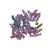
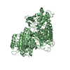
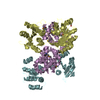
| Title | ANTI-P24 (HIV-1) FAB FRAGMENT CB41 COMPLEXED WITH AN EPITOPE-HOMOLOGOUS PEPTIDE | ||||||
 Components Components |
| ||||||
 Keywords Keywords | COMPLEX (ANTIBODY/PEPTIDE) / POLYSPECIFICITY / CROSS REACTIVITY / FAB-FRAGMENT /  HIV-1 / COMPLEX (ANTIBODY-PEPTIDE) / COMPLEX (ANTIBODY-PEPTIDE) complex HIV-1 / COMPLEX (ANTIBODY-PEPTIDE) / COMPLEX (ANTIBODY-PEPTIDE) complex | ||||||
| Function / homology |  Function and homology information Function and homology informationimmunoglobulin complex, circulating / immunoglobulin receptor binding / complement activation, classical pathway / antigen binding / antibacterial humoral response / blood microparticle / extracellular exosomeSimilarity search - Function | ||||||
| Biological species |  Mus musculus (house mouse) Mus musculus (house mouse) | ||||||
| Method | X-RAY DIFFRACTION / SYNCHROTRON / MOLECULAR REPLACEMENT / Resolution: 2.6 Å | ||||||
 Authors Authors | Keitel, T. / Kramer, A. / Wessner, H. / Scholz, C. / Schneider-Mergener, J. / Hoehne, W. | ||||||
 Citation Citation | Journal: Cell(Cambridge,Mass.) / Year: 1997 Title: Crystallographic analysis of anti-p24 (HIV-1) monoclonal antibody cross-reactivity and polyspecificity. Authors: Keitel, T. / Kramer, A. / Wessner, H. / Scholz, C. / Schneider-Mergener, J. / Hohne, W. | ||||||
| History |
|
- Structure visualization
| Structure viewer | Molecule: MolmilJmol/JSmol |
|---|
- Downloads & links
-Download
| PDBx/mmCIF format | 1bog.cif.gz | 92.5 KB | Display | PDBx/mmCIF format |
|---|---|---|---|---|
| PDB format | pdb1bog.ent.gz | 74.7 KB | Display | PDB format |
| PDBx/mmJSON format | 1bog.json.gz | Tree view | PDBx/mmJSON format | |
| Others |  Other downloads Other downloads |
-Validation report
| Arichive directory | https://data.pdbj.org/pub/pdb/validation_reports/bo/1bogftp://data.pdbj.org/pub/pdb/validation_reports/bo/1bog | HTTPS FTP |
|---|
-Related structure data
| Related structure data | 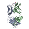 1cfnC 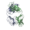 1cfqC 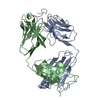 1cfsC 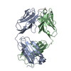 1cftC 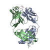 1hi6C  2hfl S: Starting model for refinement C: citing same article ( |
|---|---|
| Similar structure data |


-Links
 PDBj
PDBj


- Assembly
| Deposited unit | 
| ||||||||
|---|---|---|---|---|---|---|---|---|---|
| 1 | 
| ||||||||
| Unit cell |
|
-Components
| #1: Antibody | Mass: 23972.771 Da / Num. of mol.: 1 / Fragment: FAB FRAGMENT / Source method: isolated from a natural source / Details: FAB DERIVED FROM IGG2A KAPPA / Source: (natural) Mus musculus (house mouse) / Cell line: CB 4-1-1-F6 B-CELL HYBRIDOMA / Strain: BALB-C / References: GenBank: 387371 |
|---|---|
| #2: Antibody | Mass: 22669.508 Da / Num. of mol.: 1 / Fragment: FAB FRAGMENT / Source method: isolated from a natural source / Details: FAB DERIVED FROM IGG2A KAPPA / Source: (natural) Mus musculus (house mouse) / Cell line: CB 4/1/1/F6 B-CELL HYBRIDOMA / Strain: BALB/C / References: UniProt: P01864 |
| #3: Protein/peptide | Mass: 1186.292 Da / Num. of mol.: 1 Source method: isolated from a genetically manipulated source |
| #4: Water | ChemComp-HOH / Water Mass: 18.015 Da / Num. of mol.: 48 / Source method: isolated from a natural source / Formula: H2O Mass: 18.015 Da / Num. of mol.: 48 / Source method: isolated from a natural source / Formula: H2O |
-Experimental details
-Experiment
| Experiment | Method: X-RAY DIFFRACTION / Number of used crystals: 1 |
|---|
- Sample preparation
| Crystal | Density Matthews: 4.8 Å3/Da / Density % sol: 71 % | ||||||||||||||||||||||||||||||
|---|---|---|---|---|---|---|---|---|---|---|---|---|---|---|---|---|---|---|---|---|---|---|---|---|---|---|---|---|---|---|---|
| Crystal grow | pH: 7.5 / Details: pH 7.5 | ||||||||||||||||||||||||||||||
| Crystal grow | *PLUS Method: vapor diffusion, hanging drop | ||||||||||||||||||||||||||||||
| Components of the solutions | *PLUS
|
-Data collection
| Diffraction | Mean temperature: 288 K |
|---|---|
| Diffraction source | Source: SYNCHROTRON / Site: EMBL/DESY, HAMBURG  / Beamline: X11 / Wavelength: 0.918 / Beamline: X11 / Wavelength: 0.918 |
| Detector | Type: MARRESEARCH / Detector: IMAGE PLATE / Date: Oct 1, 1995 / Details: BENT MIRROR |
| Radiation | Monochromator: GE(111) / Monochromatic (M) / Laue (L): M / Scattering type: x-ray |
| Radiation wavelength | Wavelength: 0.918 Å / Relative weight: 1 |
| Reflection | Resolution: 2.6→91 Å / Num. obs: 30270 / % possible obs: 96 % / Observed criterion σ(I): 2 / Redundancy: 5.5 % / Biso Wilson estimate: 42 Å2 / Rmerge(I) obs: 0.076 |
- Processing
| Software |
| ||||||||||||||||||||||||||||||||||||||||||||||||||||||||||||||||||||||||||||||||||||
|---|---|---|---|---|---|---|---|---|---|---|---|---|---|---|---|---|---|---|---|---|---|---|---|---|---|---|---|---|---|---|---|---|---|---|---|---|---|---|---|---|---|---|---|---|---|---|---|---|---|---|---|---|---|---|---|---|---|---|---|---|---|---|---|---|---|---|---|---|---|---|---|---|---|---|---|---|---|---|---|---|---|---|---|---|---|
| Refinement | Method to determine structure: MOLECULAR REPLACEMENT Starting model: PDB ENTRY 2HFL 2hfl Resolution: 2.6→91 Å / Cross valid method: THROUGHOUT / σ(F): 2
| ||||||||||||||||||||||||||||||||||||||||||||||||||||||||||||||||||||||||||||||||||||
| Refinement step | Cycle: LAST / Resolution: 2.6→91 Å
| ||||||||||||||||||||||||||||||||||||||||||||||||||||||||||||||||||||||||||||||||||||
| Refine LS restraints |
| ||||||||||||||||||||||||||||||||||||||||||||||||||||||||||||||||||||||||||||||||||||
| Software | *PLUS Name: REFMAC / Classification: refinement | ||||||||||||||||||||||||||||||||||||||||||||||||||||||||||||||||||||||||||||||||||||
| Refinement | *PLUS Rfactor obs: 0.246 | ||||||||||||||||||||||||||||||||||||||||||||||||||||||||||||||||||||||||||||||||||||
| Solvent computation | *PLUS | ||||||||||||||||||||||||||||||||||||||||||||||||||||||||||||||||||||||||||||||||||||
| Displacement parameters | *PLUS |
