 Movie
Movie Controller

[English] 日本語
 Yorodumi
- PDB-1bmt: HOW A PROTEIN BINDS B12: A 3.O ANGSTROM X-RAY STRUCTURE OF THE B1... -
+ Open data

- Basic information
| Entry | Database: PDB / ID: 1bmt | ||||||
|---|---|---|---|---|---|---|---|
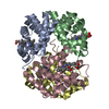
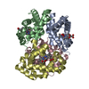
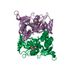
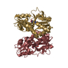
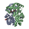
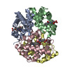
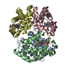
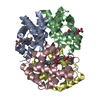
| Title | HOW A PROTEIN BINDS B12: A 3.O ANGSTROM X-RAY STRUCTURE OF THE B12-BINDING DOMAINS OF METHIONINE SYNTHASE | ||||||
 Components Components | METHIONINE SYNTHASE | ||||||
 Keywords Keywords | METHYLTRANSFERASE | ||||||
| Function / homology |  Function and homology informationmethionine synthase / methionine synthase activity / homocysteine metabolic process / methionine biosynthetic process / cobalamin binding / tetrahydrofolate metabolic process / tetrahydrofolate interconversion / methylation / zinc ion binding / cytosol / cytoplasm Function and homology informationmethionine synthase / methionine synthase activity / homocysteine metabolic process / methionine biosynthetic process / cobalamin binding / tetrahydrofolate metabolic process / tetrahydrofolate interconversion / methylation / zinc ion binding / cytosol / cytoplasmSimilarity search - Function | ||||||
| Biological species |  Escherichia coli (E. coli) Escherichia coli (E. coli) | ||||||
| Method | X-RAY DIFFRACTION / Resolution: 3 Å | ||||||
 Authors Authors | Drennan, C.L. / Huang, S. / Drummond, J.T. / Matthews, R.G. / Ludwig, M.L. | ||||||
 Citation Citation | Journal: Science / Year: 1994 Title: How a protein binds B12: A 3.0 A X-ray structure of B12-binding domains of methionine synthase. Authors: Drennan, C.L. / Huang, S. / Drummond, J.T. / Matthews, R.G. / Ludwig, M.L. #1: Journal: Curr.Opin.Struct.Biol. / Year: 1994Title: Cobalamin-Dependent Methionine Synthase: The Structure of a Methylcobalamin-Binding Fragment and Implications for Other B12-Dependent Enzymes Authors: Drennan, C.L. / Matthews, R.G. / Ludwig, M.L. | ||||||
| History |
|
- Structure visualization
| Structure viewer | Molecule: MolmilJmol/JSmol |
|---|
- Downloads & links
-Download
| PDBx/mmCIF format | 1bmt.cif.gz | 111 KB | Display | PDBx/mmCIF format |
|---|---|---|---|---|
| PDB format | pdb1bmt.ent.gz | 86 KB | Display | PDB format |
| PDBx/mmJSON format | 1bmt.json.gz | Tree view | PDBx/mmJSON format | |
| Others |  Other downloads Other downloads |
-Validation report
| Arichive directory | https://data.pdbj.org/pub/pdb/validation_reports/bm/1bmtftp://data.pdbj.org/pub/pdb/validation_reports/bm/1bmt | HTTPS FTP |
|---|
-Related structure data
| Similar structure data |
|---|


-Links
 PDBj
PDBj- Assembly
| Deposited unit | 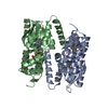
| ||||||||
|---|---|---|---|---|---|---|---|---|---|
| 1 |
| ||||||||
| Unit cell |
|
-Components
| #1: Protein | Mass: 27209.154 Da / Num. of mol.: 2 Source method: isolated from a genetically manipulated source Source: (gene. exp.) Escherichia coli (E. coli) / Production host: Escherichia coli (E. coli) / References: UniProt: P13009, methionine synthase#2: Chemical |   Mass: 1344.382 Da / Num. of mol.: 2 / Source method: obtained synthetically / Formula: C63H91CoN13O14P Mass: 1344.382 Da / Num. of mol.: 2 / Source method: obtained synthetically / Formula: C63H91CoN13O14P#3: Water | ChemComp-HOH / | Water Mass: 18.015 Da / Num. of mol.: 2 / Source method: isolated from a natural source / Formula: H2O Mass: 18.015 Da / Num. of mol.: 2 / Source method: isolated from a natural source / Formula: H2OCompound details | THE SECONDARY STRUCTURAL ELEMENTS WERE IDENTIFIED BY HYDROGEN BONDING PATTERNS AND THE METHOD OF ...THE SECONDARY STRUCTURAL | |
|---|
-Experimental details
-Experiment
| Experiment | Method: X-RAY DIFFRACTION |
|---|
- Sample preparation
| Crystal | Density Matthews: 2.55 Å3/Da / Density % sol: 51.75 % | |||||||||||||||
|---|---|---|---|---|---|---|---|---|---|---|---|---|---|---|---|---|
| Crystal grow | *PLUS Temperature: 20 ℃ / pH: 7.5 / Method: unknown | |||||||||||||||
| Components of the solutions | *PLUS
|
-Data collection
| Radiation | Scattering type: x-ray |
|---|---|
| Radiation wavelength | Relative weight: 1 |
| Reflection | *PLUS Highest resolution: 3 Å / Num. obs: 10417 / % possible obs: 89 % / Rmerge(I) obs: 0.054 |
| Reflection shell | *PLUS Mean I/σ(I) obs: 2 |
- Processing
| Software |
| ||||||||||||||||||||||||||||||||||||||||||||||||||||||||||||
|---|---|---|---|---|---|---|---|---|---|---|---|---|---|---|---|---|---|---|---|---|---|---|---|---|---|---|---|---|---|---|---|---|---|---|---|---|---|---|---|---|---|---|---|---|---|---|---|---|---|---|---|---|---|---|---|---|---|---|---|---|---|
| Refinement | Resolution: 3→8 Å / σ(F): 2 /
| ||||||||||||||||||||||||||||||||||||||||||||||||||||||||||||
| Refinement step | Cycle: LAST / Resolution: 3→8 Å
| ||||||||||||||||||||||||||||||||||||||||||||||||||||||||||||
| Refine LS restraints |
| ||||||||||||||||||||||||||||||||||||||||||||||||||||||||||||
| Refinement | *PLUS | ||||||||||||||||||||||||||||||||||||||||||||||||||||||||||||
| Solvent computation | *PLUS | ||||||||||||||||||||||||||||||||||||||||||||||||||||||||||||
| Displacement parameters | *PLUS | ||||||||||||||||||||||||||||||||||||||||||||||||||||||||||||
| Refine LS restraints | *PLUS
|
