 Movie
Movie Controller


+ Open data

- Basic information
| Entry | Database: PDB / ID: 1bh5 | ||||||
|---|---|---|---|---|---|---|---|
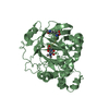
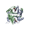
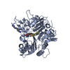
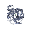
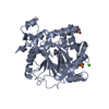
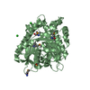
| Title | HUMAN GLYOXALASE I Q33E, E172Q DOUBLE MUTANT | ||||||
 Components Components | LACTOYLGLUTATHIONE LYASE | ||||||
 Keywords Keywords | LYASE / LACTOYLGLUTATHIONE LYASE / GLYOXALASE I | ||||||
| Function / homology |  Function and homology informationlactoylglutathione lyase / lactoylglutathione lyase activity / methylglyoxal metabolic process / Pyruvate metabolism / glutathione metabolic process / osteoclast differentiation / carbohydrate metabolic process / regulation of transcription by RNA polymerase II / negative regulation of apoptotic process / extracellular exosome ...lactoylglutathione lyase / lactoylglutathione lyase activity / methylglyoxal metabolic process / Pyruvate metabolism / glutathione metabolic process / osteoclast differentiation / carbohydrate metabolic process / regulation of transcription by RNA polymerase II / negative regulation of apoptotic process / extracellular exosome / zinc ion binding / nucleoplasm / plasma membrane / cytosol / cytoplasm Function and homology informationlactoylglutathione lyase / lactoylglutathione lyase activity / methylglyoxal metabolic process / Pyruvate metabolism / glutathione metabolic process / osteoclast differentiation / carbohydrate metabolic process / regulation of transcription by RNA polymerase II / negative regulation of apoptotic process / extracellular exosome ...lactoylglutathione lyase / lactoylglutathione lyase activity / methylglyoxal metabolic process / Pyruvate metabolism / glutathione metabolic process / osteoclast differentiation / carbohydrate metabolic process / regulation of transcription by RNA polymerase II / negative regulation of apoptotic process / extracellular exosome / zinc ion binding / nucleoplasm / plasma membrane / cytosol / cytoplasmSimilarity search - Function | ||||||
| Biological species |  Homo sapiens (human) Homo sapiens (human) | ||||||
| Method | X-RAY DIFFRACTION / molecular replacement / Resolution: 2.2 Å | ||||||
 Authors Authors | Cameron, A.D. / Jones, T.A. | ||||||
 Citation Citation | Journal: J.Biol.Chem. / Year: 1998 Title: Involvement of an active-site Zn2+ ligand in the catalytic mechanism of human glyoxalase I. Authors: Ridderstrom, M. / Cameron, A.D. / Jones, T.A. / Mannervik, B. | ||||||
| History |
|
- Structure visualization
| Structure viewer | Molecule: MolmilJmol/JSmol |
|---|
- Downloads & links
-Download
| PDBx/mmCIF format | 1bh5.cif.gz | 166.4 KB | Display | PDBx/mmCIF format |
|---|---|---|---|---|
| PDB format | pdb1bh5.ent.gz | 132.3 KB | Display | PDB format |
| PDBx/mmJSON format | 1bh5.json.gz | Tree view | PDBx/mmJSON format | |
| Others |  Other downloads Other downloads |
-Validation report
| Arichive directory | https://data.pdbj.org/pub/pdb/validation_reports/bh/1bh5ftp://data.pdbj.org/pub/pdb/validation_reports/bh/1bh5 | HTTPS FTP |
|---|
-Related structure data
| Related structure data | 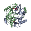 1froS S: Starting model for refinement |
|---|---|
| Similar structure data |


-Links
 PDBj
PDBj- Assembly
| Deposited unit | 
| ||||||||||||||||
|---|---|---|---|---|---|---|---|---|---|---|---|---|---|---|---|---|---|
| 1 | 
| ||||||||||||||||
| 2 | 
| ||||||||||||||||
| Unit cell |
| ||||||||||||||||
| Noncrystallographic symmetry (NCS) | NCS oper:
| ||||||||||||||||
| Details | THE BIOLOGICALLY ACTIVE MOLECULE IS THE DIMER (MOLECULES A AND B OR C AND D). THE TWO DIMERS IN THE ASYMMETRIC UNIT ARE SITUATED IN SIMILAR CRYSTALLOGRAHIC ENVIRONMENTS. DURING REFINEMENT NON-CRYSTALLOGRAPHIC RESTRAINTS WERE APPLIED BETWEEN THE A AND C MOLECULES AND BETWEEN THE B AND D MOLECULES). NO NCS RESTRAINTS WERE APPLIED BETWEEN THE TWO MOLECULES OF THE DIMERS. DISORDERED SIDE CHAINS HAVE BEEN INCLUDED WITH OCCUPANCIES OF 0.01. RESIDUE CYS 60 IN THE B AND D MOLECULES APPEARS TO BE INVOLVED IN A DISULFIDE BRIDGE WITH 2-MERCAPTOETHANOL AS STATED FOR 1FRO.PDB. THE 2-MERCAPTOETHANOL HAS NOT BEEN MODELLED DUE TO THE LIMITED NUMBER OF DATA. |
-Components
| #1: Protein | / GLYOXALASE I Mass: 20672.520 Da / Num. of mol.: 4 / Mutation: Q33E, E172Q Source method: isolated from a genetically manipulated source Source: (gene. exp.) Homo sapiens (human) / Plasmid: PKK223-3 / Production host:  Escherichia coli (E. coli) / References: UniProt: Q04760, lactoylglutathione lyase Escherichia coli (E. coli) / References: UniProt: Q04760, lactoylglutathione lyase#2: Chemical | ChemComp-ZN /   Mass: 65.409 Da / Num. of mol.: 4 / Source method: obtained synthetically / Formula: Zn Mass: 65.409 Da / Num. of mol.: 4 / Source method: obtained synthetically / Formula: Zn#3: Chemical | ChemComp-GTX /   Mass: 392.491 Da / Num. of mol.: 4 / Source method: obtained synthetically / Formula: C16H30N3O6S Mass: 392.491 Da / Num. of mol.: 4 / Source method: obtained synthetically / Formula: C16H30N3O6S#4: Water | ChemComp-HOH / | Water Mass: 18.015 Da / Num. of mol.: 507 / Source method: isolated from a natural source / Formula: H2O Mass: 18.015 Da / Num. of mol.: 507 / Source method: isolated from a natural source / Formula: H2O |
|---|
-Experimental details
-Experiment
| Experiment | Method: X-RAY DIFFRACTION / Number of used crystals: 1 |
|---|
- Sample preparation
| Crystal | Density Matthews: 2 Å3/Da / Density % sol: 42 % | ||||||||||||||||||||||||||||||||||||||||||||||||||||||||||||
|---|---|---|---|---|---|---|---|---|---|---|---|---|---|---|---|---|---|---|---|---|---|---|---|---|---|---|---|---|---|---|---|---|---|---|---|---|---|---|---|---|---|---|---|---|---|---|---|---|---|---|---|---|---|---|---|---|---|---|---|---|---|
| Crystal grow | pH: 5.8 Details: PROTEIN WAS CRYSTALLISED FROM PEG 2000 MONOMETHLY ETHER 50 MM MES PH 5.8, 0.1M NACL | ||||||||||||||||||||||||||||||||||||||||||||||||||||||||||||
| Crystal grow | *PLUS Temperature: 15 ℃ / Method: vapor diffusion, hanging drop / Details: Cameron, A.D., (1997) EMBO J., 16, 3386. | ||||||||||||||||||||||||||||||||||||||||||||||||||||||||||||
| Components of the solutions | *PLUS
|
-Data collection
| Diffraction | Mean temperature: 100 K |
|---|---|
| Diffraction source | Source: ROTATING ANODE / Type: RIGAKU / Wavelength: 1.5418 |
| Detector | Type: RIGAKU RAXIS / Detector: IMAGE PLATE / Date: Mar 23, 1997 |
| Radiation | Monochromator: GRAPHITE(002) / Monochromatic (M) / Laue (L): M / Scattering type: x-ray |
| Radiation wavelength | Wavelength: 1.5418 Å / Relative weight: 1 |
| Reflection | Resolution: 2.2→30 Å / Num. obs: 33195 / % possible obs: 89.7 % / Redundancy: 3.5 % / Biso Wilson estimate: 13 Å2 / Rmerge(I) obs: 0.048 / Net I/σ(I): 27 |
| Reflection shell | Resolution: 2.2→2.32 Å / Redundancy: 1.8 % / Rmerge(I) obs: 0.128 / Mean I/σ(I) obs: 5.5 / % possible all: 63.1 |
| Reflection shell | *PLUS % possible obs: 63 % |
- Processing
| Software |
| ||||||||||||||||||||||||||||||||||||||||||||||||||||||||||||||||||||||||||||||||||||
|---|---|---|---|---|---|---|---|---|---|---|---|---|---|---|---|---|---|---|---|---|---|---|---|---|---|---|---|---|---|---|---|---|---|---|---|---|---|---|---|---|---|---|---|---|---|---|---|---|---|---|---|---|---|---|---|---|---|---|---|---|---|---|---|---|---|---|---|---|---|---|---|---|---|---|---|---|---|---|---|---|---|---|---|---|---|
| Refinement | Method to determine structure: molecular replacement Starting model: PDB ENTRY 1FRO Resolution: 2.2→30 Å / Cross valid method: THROUGHOUT
| ||||||||||||||||||||||||||||||||||||||||||||||||||||||||||||||||||||||||||||||||||||
| Displacement parameters | Biso mean: 16 Å2 | ||||||||||||||||||||||||||||||||||||||||||||||||||||||||||||||||||||||||||||||||||||
| Refine analyze | Luzzati sigma a obs: 0.26 Å | ||||||||||||||||||||||||||||||||||||||||||||||||||||||||||||||||||||||||||||||||||||
| Refinement step | Cycle: LAST / Resolution: 2.2→30 Å
| ||||||||||||||||||||||||||||||||||||||||||||||||||||||||||||||||||||||||||||||||||||
| Refine LS restraints |
| ||||||||||||||||||||||||||||||||||||||||||||||||||||||||||||||||||||||||||||||||||||
| Software | *PLUS Name: REFMAC / Classification: refinement | ||||||||||||||||||||||||||||||||||||||||||||||||||||||||||||||||||||||||||||||||||||
| Refinement | *PLUS Rfactor obs: 0.25 / Rfactor Rfree: 0.28 | ||||||||||||||||||||||||||||||||||||||||||||||||||||||||||||||||||||||||||||||||||||
| Solvent computation | *PLUS | ||||||||||||||||||||||||||||||||||||||||||||||||||||||||||||||||||||||||||||||||||||
| Displacement parameters | *PLUS |
