 Movie
Movie Controller

[English] 日本語
 Yorodumi
- PDB-1bdx: E. COLI DNA HELICASE RUVA WITH BOUND DNA HOLLIDAY JUNCTION, ALPHA... -
+ Open data

- Basic information
| Entry | Database: PDB / ID: 1bdx | ||||||
|---|---|---|---|---|---|---|---|
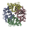
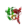
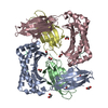
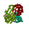
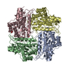
| Title | E. COLI DNA HELICASE RUVA WITH BOUND DNA HOLLIDAY JUNCTION, ALPHA CARBONS AND PHOSPHATE ATOMS ONLY | ||||||
 Components Components |
| ||||||
 Keywords Keywords | TRANSFERASE/DNA / DNA-BINDING /  BRANCH MIGRATION / HOLLIDAY JUNCTION / RUV / COMPLEX DNA-BINDING PROTEIN-DNA / TRANSFERASE-DNA COMPLEX BRANCH MIGRATION / HOLLIDAY JUNCTION / RUV / COMPLEX DNA-BINDING PROTEIN-DNA / TRANSFERASE-DNA COMPLEX | ||||||
| Function / homology |  Function and homology information Function and homology informationHolliday junction helicase complex / Holliday junction resolvase complex / four-way junction helicase activity / recombinational repair / SOS response / four-way junction DNA binding / response to radiation / ATP binding / identical protein binding / cytoplasmSimilarity search - Function | ||||||
| Biological species |  Escherichia coli BL21 (bacteria) Escherichia coli BL21 (bacteria) | ||||||
| Method | X-RAY DIFFRACTION / SYNCHROTRON / MOLECULAR REPLACEMENT, MIR / Resolution: 6 Å | ||||||
 Authors Authors | Hargreaves, D. / Rice, D.W. / Sedelnikova, S.E. / Artymiuk, P.J. / Lloyd, R.G. / Rafferty, J.B. | ||||||
 Citation Citation | Journal: Nat.Struct.Biol. / Year: 1998 Title: Crystal structure of E.coli RuvA with bound DNA Holliday junction at 6 A resolution. Authors: Hargreaves, D. / Rice, D.W. / Sedelnikova, S.E. / Artymiuk, P.J. / Lloyd, R.G. / Rafferty, J.B. #1: Journal: Science / Year: 1996Title: Crystal Structure of DNA Recombination Protein Ruva and a Model for its Binding to the Holliday Junction Authors: Rafferty, J.B. / Sedelnikova, S.E. / Hargreaves, D. / Artymiuk, P.J. / Baker, P.J. / Sharples, G.J. / Mahdi, A.A. / Lloyd, R.G. / Rice, D.W. | ||||||
| History |
|
- Structure visualization
| Structure viewer | Molecule: MolmilJmol/JSmol |
|---|
- Downloads & links
-Download
| PDBx/mmCIF format | 1bdx.cif.gz | 35.8 KB | Display | PDBx/mmCIF format |
|---|---|---|---|---|
| PDB format | pdb1bdx.ent.gz | 19.8 KB | Display | PDB format |
| PDBx/mmJSON format | 1bdx.json.gz | Tree view | PDBx/mmJSON format | |
| Others |  Other downloads Other downloads |
-Validation report
| Arichive directory | https://data.pdbj.org/pub/pdb/validation_reports/bd/1bdxftp://data.pdbj.org/pub/pdb/validation_reports/bd/1bdx | HTTPS FTP |
|---|
-Related structure data
| Related structure data | 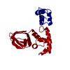 1cukS S: Starting model for refinement |
|---|---|
| Similar structure data |

-Links
 PDBj
PDBj


- Assembly
| Deposited unit | 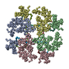
| ||||||||||||||||
|---|---|---|---|---|---|---|---|---|---|---|---|---|---|---|---|---|---|
| 1 |
| ||||||||||||||||
| Unit cell |
| ||||||||||||||||
| Noncrystallographic symmetry (NCS) | NCS oper:
|
-Components
| #1: DNA chain | Mass: 4898.191 Da / Num. of mol.: 4 / Source method: obtained synthetically #2: Protein | Mass: 22111.668 Da / Num. of mol.: 4 Source method: isolated from a genetically manipulated source Source: (gene. exp.) Escherichia coli BL21(DE3) (bacteria) / Species: Escherichia coli / Strain: BL21List of strains of Escherichia coli / Variant: DE3 / Plasmid: PAM159 / Species (production host): Escherichia coli / Gene (production host): RUVA / Production host: Escherichia coli BL21(DE3) (bacteria) / Strain (production host): BL21 / Variant (production host): DE3 / References: UniProt: P0A809 |
|---|
-Experimental details
-Experiment
| Experiment | Method: X-RAY DIFFRACTION / Number of used crystals: 1 |
|---|
- Sample preparation
| Crystal | Density Matthews: 4.46 Å3/Da / Density % sol: 48 % Description: MOLECULAR REPLACEMENT PHASES WERE ONLY GOOD ENOUGH TO USE IN LOCATING HEAVY ATOMS BY DIFFERENCE FOURIER AND WERE THEN ABANDONED IN FAVOUR OF MIR PHASES. | |||||||||||||||||||||||||
|---|---|---|---|---|---|---|---|---|---|---|---|---|---|---|---|---|---|---|---|---|---|---|---|---|---|---|
| Crystal grow | Method: vapor diffusion, hanging drop / pH: 6.5 Details: PROTEIN/DNA COMPLEX WAS CRYSTALLISED FROM 0.85M SODIUM ACETATE BUFFERED WITH 100MM IMIDAZOLE AT PH 6.5, VAPOR DIFFUSION, HANGING DROP | |||||||||||||||||||||||||
| Components of the solutions |
| |||||||||||||||||||||||||
| Crystal grow | *PLUS Details: Hargreaves, D., (1999) Acta. Crystallogr., D55, 263.PH range low: 7.5 / PH range high: 6.5 | |||||||||||||||||||||||||
| Components of the solutions | *PLUS
|
-Data collection
| Diffraction | Mean temperature: 100 K |
|---|---|
| Diffraction source | Source: SYNCHROTRON / Site: SRS  / Beamline: PX9.6 / Wavelength: 0.87 / Beamline: PX9.6 / Wavelength: 0.87 |
| Detector | Type: MARRESEARCH / Detector: IMAGE PLATE / Date: Dec 15, 1997 / Details: MIRRORS |
| Radiation | Monochromator: SI CRYSTAL / Protocol: SINGLE WAVELENGTH / Monochromatic (M) / Laue (L): M / Scattering type: x-ray |
| Radiation wavelength | Wavelength: 0.87 Å / Relative weight: 1 |
| Reflection | Resolution: 5.7→17.6 Å / Num. obs: 5263 / % possible obs: 94 % / Observed criterion σ(I): 2 / Redundancy: 2.1 % / Rmerge(I) obs: 0.043 / Rsym value: 0.043 / Net I/σ(I): 6.8 |
| Reflection shell | Resolution: 5.7→6.01 Å / Redundancy: 2.1 % / Rmerge(I) obs: 0.224 / Mean I/σ(I) obs: 3.3 / Rsym value: 0.224 / % possible all: 94.2 |
| Reflection | *PLUS % possible obs: 94 % / Num. measured all: 10906 |
- Processing
| Software |
| |||||||||||||||||||||||||||
|---|---|---|---|---|---|---|---|---|---|---|---|---|---|---|---|---|---|---|---|---|---|---|---|---|---|---|---|---|
| Refinement | Method to determine structure: MOLECULAR REPLACEMENT, MIR Starting model: 1CUK Highest resolution: 6 Å Details: OWING TO THE LOW RESOLUTION OF THE DATA, NO POSITIONAL REFINEMENT OF THE PROTEIN RESIDUES OR DNA WAS PERFORMED | |||||||||||||||||||||||||||
| Refinement step | Cycle: LAST / Highest resolution: 6 Å
|
