 Movie
Movie Controller

[English] 日本語
 Yorodumi
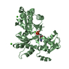
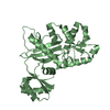
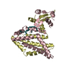
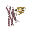
- PDB-1bc5: CHEMOTAXIS RECEPTOR RECOGNITION BY PROTEIN METHYLTRANSFERASE CHER -
+ Open data

- Basic information
| Entry | Database: PDB / ID: 1bc5 | ||||||
|---|---|---|---|---|---|---|---|
| Title | CHEMOTAXIS RECEPTOR RECOGNITION BY PROTEIN METHYLTRANSFERASE CHER | ||||||
 Components Components |
| ||||||
 Keywords Keywords | COMPLEX (METHYLTRANSFERASE/PEPTIDE) /  METHYLTRANSFERASE / PEPTIDE BINDING / CHEMOTAXIS RECEPTOR / COMPLEX (METHYLTRANSFERASE-PEPTIDE) / COMPLEX (METHYLTRANSFERASE-PEPTIDE) complex METHYLTRANSFERASE / PEPTIDE BINDING / CHEMOTAXIS RECEPTOR / COMPLEX (METHYLTRANSFERASE-PEPTIDE) / COMPLEX (METHYLTRANSFERASE-PEPTIDE) complex | ||||||
| Function / homology |  Function and homology informationprotein-glutamate O-methyltransferase / protein-glutamate O-methyltransferase activity / chemotaxis / methylation Function and homology informationprotein-glutamate O-methyltransferase / protein-glutamate O-methyltransferase activity / chemotaxis / methylationSimilarity search - Function | ||||||
| Biological species |  Salmonella typhimurium (bacteria) Salmonella typhimurium (bacteria) | ||||||
| Method | X-RAY DIFFRACTION / MOLECULAR REPLACEMENT / Resolution: 2.2 Å | ||||||
 Authors Authors | Djordjevic, S. / Stock, A.M. | ||||||
 Citation Citation | Journal: Nat.Struct.Biol. / Year: 1998 Title: Chemotaxis receptor recognition by protein methyltransferase CheR. Authors: Djordjevic, S. / Stock, A.M. | ||||||
| History |
|
- Structure visualization
| Structure viewer | Molecule: MolmilJmol/JSmol |
|---|
- Downloads & links
-Download
| PDBx/mmCIF format | 1bc5.cif.gz | 67.8 KB | Display | PDBx/mmCIF format |
|---|---|---|---|---|
| PDB format | pdb1bc5.ent.gz | 52.7 KB | Display | PDB format |
| PDBx/mmJSON format | 1bc5.json.gz | Tree view | PDBx/mmJSON format | |
| Others |  Other downloads Other downloads |
-Validation report
| Arichive directory | https://data.pdbj.org/pub/pdb/validation_reports/bc/1bc5ftp://data.pdbj.org/pub/pdb/validation_reports/bc/1bc5 | HTTPS FTP |
|---|
-Related structure data
| Related structure data |  1af7S S: Starting model for refinement |
|---|---|
| Similar structure data |


-Links
 PDBj
PDBj
- Assembly
| Deposited unit | 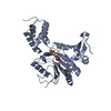
| ||||||||
|---|---|---|---|---|---|---|---|---|---|
| 1 |
| ||||||||
| Unit cell |
|
-Components
| #1: Protein | Mass: 30992.387 Da / Num. of mol.: 1 Source method: isolated from a genetically manipulated source Source: (gene. exp.) Salmonella typhimurium (bacteria) / Plasmid: PME43 / Production host: Escherichia coli (E. coli) / Strain (production host): HB101References: UniProt: P07801, protein-glutamate O-methyltransferase |
|---|---|
| #2: Protein/peptide | Mass: 721.757 Da / Num. of mol.: 1 Fragment: C-TERMINAL PENTAPEPTIDE, ACETYLATED ASN-TRP-GLU-THR-PHE Source method: isolated from a genetically manipulated source |
| #3: Chemical | ChemComp-CO /   Mass: 58.933 Da / Num. of mol.: 1 / Source method: obtained synthetically / Formula: Co Mass: 58.933 Da / Num. of mol.: 1 / Source method: obtained synthetically / Formula: Co |
| #4: Chemical | ChemComp-SAH / S-Adenosyl-L-homocysteine  Type: L-peptide linking / Mass: 384.411 Da / Num. of mol.: 1 / Source method: obtained synthetically / Formula: C14H20N6O5S Type: L-peptide linking / Mass: 384.411 Da / Num. of mol.: 1 / Source method: obtained synthetically / Formula: C14H20N6O5S |
| #5: Water | ChemComp-HOH / Water Mass: 18.015 Da / Num. of mol.: 121 / Source method: isolated from a natural source / Formula: H2O Mass: 18.015 Da / Num. of mol.: 121 / Source method: isolated from a natural source / Formula: H2O |
-Experimental details
-Experiment
| Experiment | Method: X-RAY DIFFRACTION / Number of used crystals: 1 |
|---|
- Sample preparation
| Crystal | Density Matthews: 2.55 Å3/Da / Density % sol: 52 % | ||||||||||||||||||||||||||||||||||||||||||||||||||||||||||||
|---|---|---|---|---|---|---|---|---|---|---|---|---|---|---|---|---|---|---|---|---|---|---|---|---|---|---|---|---|---|---|---|---|---|---|---|---|---|---|---|---|---|---|---|---|---|---|---|---|---|---|---|---|---|---|---|---|---|---|---|---|---|
| Crystal grow | pH: 6.8 Details: PROTEIN WAS CRYSTALLIZED FROM 30 % PEG 5000 MME, 25 MM IMIDAZOLE PH 6.8, 15 MM COCL2, 1MM BETA-MERCAPTOETHANOL. | ||||||||||||||||||||||||||||||||||||||||||||||||||||||||||||
| Crystal grow | *PLUS Method: vapor diffusion, sitting drop | ||||||||||||||||||||||||||||||||||||||||||||||||||||||||||||
| Components of the solutions | *PLUS
|
-Data collection
| Diffraction | Mean temperature: 263 K |
|---|---|
| Diffraction source | Source: ROTATING ANODE / Type: RIGAKU RUH2R / Wavelength: 1.5418 |
| Detector | Type: RIGAKU / Detector: IMAGE PLATE / Date: Nov 1, 1997 / Details: MIRRORS |
| Radiation | Monochromator: NI FILTER / Monochromatic (M) / Laue (L): M / Scattering type: x-ray |
| Radiation wavelength | Wavelength: 1.5418 Å / Relative weight: 1 |
| Reflection | Resolution: 2.2→23 Å / Num. obs: 16682 / % possible obs: 94.5 % / Observed criterion σ(I): 2 / Redundancy: 3.1 % / Biso Wilson estimate: 35.3 Å2 / Rsym value: 0.064 / Net I/σ(I): 7.8 |
| Reflection shell | Resolution: 2.2→2.26 Å / Redundancy: 2.6 % / Mean I/σ(I) obs: 2.6 / Rsym value: 0.28 / % possible all: 89 |
| Reflection | *PLUS Num. measured all: 52170 / Rmerge(I) obs: 0.064 |
| Reflection shell | *PLUS % possible obs: 89 % / Rmerge(I) obs: 0.28 |
- Processing
| Software |
| ||||||||||||||||||||||||||||||||||||||||||||||||||||||||||||
|---|---|---|---|---|---|---|---|---|---|---|---|---|---|---|---|---|---|---|---|---|---|---|---|---|---|---|---|---|---|---|---|---|---|---|---|---|---|---|---|---|---|---|---|---|---|---|---|---|---|---|---|---|---|---|---|---|---|---|---|---|---|
| Refinement | Method to determine structure: MOLECULAR REPLACEMENT Starting model: PDB ENTRY 1AF7 Resolution: 2.2→8 Å / Data cutoff high absF: 10000000 / Isotropic thermal model: RESTRAINED / Cross valid method: THROUGHOUT / σ(F): 2
| ||||||||||||||||||||||||||||||||||||||||||||||||||||||||||||
| Displacement parameters | Biso mean: 25.3 Å2 | ||||||||||||||||||||||||||||||||||||||||||||||||||||||||||||
| Refinement step | Cycle: LAST / Resolution: 2.2→8 Å
| ||||||||||||||||||||||||||||||||||||||||||||||||||||||||||||
| Refine LS restraints |
| ||||||||||||||||||||||||||||||||||||||||||||||||||||||||||||
| Xplor file |
| ||||||||||||||||||||||||||||||||||||||||||||||||||||||||||||
| Software | *PLUS Name: X-PLOR / Version: 3.1 / Classification: refinement | ||||||||||||||||||||||||||||||||||||||||||||||||||||||||||||
| Refinement | *PLUS Rfactor Rwork: 0.204 / % reflection Rfree: 5 % / Rfactor obs: 0.204 / Rfactor Rfree: 0.288 | ||||||||||||||||||||||||||||||||||||||||||||||||||||||||||||
| Solvent computation | *PLUS | ||||||||||||||||||||||||||||||||||||||||||||||||||||||||||||
| Displacement parameters | *PLUS | ||||||||||||||||||||||||||||||||||||||||||||||||||||||||||||
| Refine LS restraints | *PLUS
|
