 Movie
Movie Controller


+ Open data

- Basic information
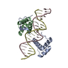
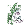
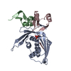
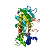
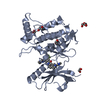
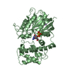
| Entry | Database: PDB / ID: 1b72 | ||||||
|---|---|---|---|---|---|---|---|
| Title | PBX1, HOMEOBOX PROTEIN HOX-B1/DNA TERNARY COMPLEX | ||||||
 Components Components |
| ||||||
 Keywords Keywords | PROTEIN/DNA /  HOMEODOMAIN / DNA / COMPLEX / DNA-BINDING PROTEIN / PROTEIN-DNA COMPLEX HOMEODOMAIN / DNA / COMPLEX / DNA-BINDING PROTEIN / PROTEIN-DNA COMPLEX | ||||||
| Function / homology |  Function and homology information Function and homology informationrhombomere 4 development / rhombomere 5 development / facial nucleus development / anatomical structure formation involved in morphogenesis / urogenital system development / facial nerve structural organization / embryonic skeletal system morphogenesis / natural killer cell differentiation / proximal/distal pattern formation / embryonic skeletal system development ...rhombomere 4 development / rhombomere 5 development / facial nucleus development / anatomical structure formation involved in morphogenesis / urogenital system development / facial nerve structural organization / embryonic skeletal system morphogenesis / natural killer cell differentiation / proximal/distal pattern formation / embryonic skeletal system development / Transcriptional regulation of pluripotent stem cells / sex differentiation / multicellular organism development / chromatin => GO:0000785 / eye development / pattern specification process / steroid biosynthetic process / embryonic limb morphogenesis / embryonic hemopoiesis / NOTCH3 Intracellular Domain Regulates Transcription / anterior/posterior pattern specification / branching involved in ureteric bud morphogenesis / adrenal gland development / positive regulation of stem cell proliferation / negative regulation of neuron differentiation / regulation of ossification / neuron development / embryonic organ development / spleen development / positive regulation of G2/M transition of mitotic cell cycle / transcription corepressor binding / thymus development / stem cell proliferation / transcription coregulator binding / RNA polymerase II transcription regulatory region sequence-specific DNA binding / animal organ morphogenesis / brain development / negative regulation of DNA-binding transcription factor activity / Activation of anterior HOX genes in hindbrain development during early embryogenesis / RNA polymerase II transcription regulator complex / G2/M transition of mitotic cell cycle / sequence-specific double-stranded DNA binding / DNA-binding transcription activator activity, RNA polymerase II-specific / DNA-binding transcription factor binding / transcription cis-regulatory region binding / DNA-binding transcription factor activity, RNA polymerase II-specific / RNA polymerase II cis-regulatory region sequence-specific DNA binding / DNA-binding transcription factor activity / protein domain specific binding / chromatin / regulation of DNA-templated transcription / regulation of transcription by RNA polymerase II / positive regulation of transcription by RNA polymerase II / DNA binding / nucleoplasm / nucleus / cytoplasmSimilarity search - Function | ||||||
| Biological species |  Homo sapiens (human) Homo sapiens (human) | ||||||
| Method | X-RAY DIFFRACTION / SYNCHROTRON / MIR / Resolution: 2.35 Å | ||||||
 Authors Authors | Piper, D.E. / Batchelor, A.H. / Chang, C.-P. / Cleary, M.L. / Wolberger, C. | ||||||
 Citation Citation | Journal: Cell(Cambridge,Mass.) / Year: 1999 Title: Structure of a HoxB1-Pbx1 heterodimer bound to DNA: role of the hexapeptide and a fourth homeodomain helix in complex formation. Authors: Piper, D.E. / Batchelor, A.H. / Chang, C.P. / Cleary, M.L. / Wolberger, C. | ||||||
| History |
|
- Structure visualization
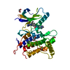
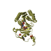
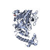
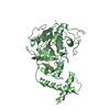
| Structure viewer | Molecule: MolmilJmol/JSmol |
|---|
- Downloads & links
-Download
| PDBx/mmCIF format | 1b72.cif.gz | 66.2 KB | Display | PDBx/mmCIF format |
|---|---|---|---|---|
| PDB format | pdb1b72.ent.gz | 45.2 KB | Display | PDB format |
| PDBx/mmJSON format | 1b72.json.gz | Tree view | PDBx/mmJSON format | |
| Others |  Other downloads Other downloads |
-Validation report
| Arichive directory | https://data.pdbj.org/pub/pdb/validation_reports/b7/1b72ftp://data.pdbj.org/pub/pdb/validation_reports/b7/1b72 | HTTPS FTP |
|---|
-Related structure data
| Similar structure data |
|---|
-Links
 PDBj
PDBj


- Assembly
| Deposited unit | 
| ||||||||||
|---|---|---|---|---|---|---|---|---|---|---|---|
| 1 |
| ||||||||||
| Unit cell |
|
-Components
| #1: DNA chain | Mass: 6139.976 Da / Num. of mol.: 1 / Source method: obtained synthetically |
|---|---|
| #2: DNA chain | Mass: 6126.994 Da / Num. of mol.: 1 / Source method: obtained synthetically |
| #3: Protein | Mass: 11443.128 Da / Num. of mol.: 1 / Fragment: HEXAPEPTIDE, HOMEODOMAIN / Mutation: 1 RESIDUE (MET) ADDED TO N-TERMINUS Source method: isolated from a genetically manipulated source Source: (gene. exp.) Homo sapiens (human) / Gene: HOXB-1 / Plasmid: PET11-D T7 PROMOTER / Gene (production host): HOXB-1 / Production host:  Escherichia coli (E. coli) / Strain (production host): BL21 (DE3) PLYSS / References: UniProt: P14653 Escherichia coli (E. coli) / Strain (production host): BL21 (DE3) PLYSS / References: UniProt: P14653 |
| #4: Protein | Mass: 10073.342 Da / Num. of mol.: 1 / Fragment: HOMEODOMAIN Source method: isolated from a genetically manipulated source Source: (gene. exp.) Homo sapiens (human) / Gene: PBX1 / Plasmid: PET11-D T7 PROMOTER / Gene (production host): PBX1 / Production host: Escherichia coli (E. coli) / Strain (production host): BL21 (DE3) PLYSS / References: UniProt: P40424 |
| #5: Water | ChemComp-HOH / Water Mass: 18.015 Da / Num. of mol.: 61 / Source method: isolated from a natural source / Formula: H2O Mass: 18.015 Da / Num. of mol.: 61 / Source method: isolated from a natural source / Formula: H2O |
-Experimental details
-Experiment
| Experiment | Method: X-RAY DIFFRACTION / Number of used crystals: 1 |
|---|
- Sample preparation
| Crystal | Density Matthews: 2.3 Å3/Da / Density % sol: 47 % | |||||||||||||||||||||||||||||||||||||||||||||||||||||||||||||||
|---|---|---|---|---|---|---|---|---|---|---|---|---|---|---|---|---|---|---|---|---|---|---|---|---|---|---|---|---|---|---|---|---|---|---|---|---|---|---|---|---|---|---|---|---|---|---|---|---|---|---|---|---|---|---|---|---|---|---|---|---|---|---|---|---|
| Crystal grow | pH: 8 / Details: pH 8.0 | |||||||||||||||||||||||||||||||||||||||||||||||||||||||||||||||
| Components of the solutions |
| |||||||||||||||||||||||||||||||||||||||||||||||||||||||||||||||
| Crystal grow | *PLUS Method: vapor diffusion, hanging dropDetails: equal volume of protein and reservoir solution were used for the drop | |||||||||||||||||||||||||||||||||||||||||||||||||||||||||||||||
| Components of the solutions | *PLUS
|
-Data collection
| Diffraction | Mean temperature: 100 K |
|---|---|
| Diffraction source | Source: SYNCHROTRON / Site: NSLS  / Beamline: X4A / Wavelength: 0.9795 / Beamline: X4A / Wavelength: 0.9795 |
| Detector | Type: RIGAKU RAXIS IV / Detector: IMAGE PLATE / Date: Oct 15, 1998 / Details: SPHERICAL RH COATED |
| Radiation | Monochromator: SI(111) / Protocol: SINGLE WAVELENGTH / Monochromatic (M) / Laue (L): M / Scattering type: x-ray |
| Radiation wavelength | Wavelength: 0.9795 Å / Relative weight: 1 |
| Reflection | Resolution: 2.35→30 Å / Num. obs: 13735 / % possible obs: 99.7 % / Observed criterion σ(I): -3 / Redundancy: 5.7 % / Biso Wilson estimate: 44 Å2 / Rsym value: 0.033 / Net I/σ(I): 27.2 |
| Reflection shell | Resolution: 2.35→2.43 Å / Redundancy: 5.6 % / Mean I/σ(I) obs: 7.7 / Rsym value: 0.248 / % possible all: 99.7 |
| Reflection | *PLUS Rmerge(I) obs: 0.033 |
| Reflection shell | *PLUS % possible obs: 99.7 % / Rmerge(I) obs: 0.248 |
- Processing
| Software |
| ||||||||||||||||||||||||||||||||||||||||||||||||||||||||||||||||||||||||||||||||
|---|---|---|---|---|---|---|---|---|---|---|---|---|---|---|---|---|---|---|---|---|---|---|---|---|---|---|---|---|---|---|---|---|---|---|---|---|---|---|---|---|---|---|---|---|---|---|---|---|---|---|---|---|---|---|---|---|---|---|---|---|---|---|---|---|---|---|---|---|---|---|---|---|---|---|---|---|---|---|---|---|---|
| Refinement | Method to determine structure: MIR / Resolution: 2.35→30 Å / Rfactor Rfree error: 0.007 / Data cutoff high rms absF: 1033240 / Isotropic thermal model: RESTRAINED / Cross valid method: THROUGHOUT / σ(F): 0 / Details: BULK SOLVENT MODEL USED
| ||||||||||||||||||||||||||||||||||||||||||||||||||||||||||||||||||||||||||||||||
| Displacement parameters | Biso mean: 53.2 Å2
| ||||||||||||||||||||||||||||||||||||||||||||||||||||||||||||||||||||||||||||||||
| Refine analyze |
| ||||||||||||||||||||||||||||||||||||||||||||||||||||||||||||||||||||||||||||||||
| Refinement step | Cycle: LAST / Resolution: 2.35→30 Å
| ||||||||||||||||||||||||||||||||||||||||||||||||||||||||||||||||||||||||||||||||
| Refine LS restraints |
| ||||||||||||||||||||||||||||||||||||||||||||||||||||||||||||||||||||||||||||||||
| LS refinement shell | Resolution: 2.35→2.5 Å / Rfactor Rfree error: 0.026 / Total num. of bins used: 6
| ||||||||||||||||||||||||||||||||||||||||||||||||||||||||||||||||||||||||||||||||
| Xplor file |
| ||||||||||||||||||||||||||||||||||||||||||||||||||||||||||||||||||||||||||||||||
| Software | *PLUS Name: CNS / Version: 0.3 / Classification: refinement | ||||||||||||||||||||||||||||||||||||||||||||||||||||||||||||||||||||||||||||||||
| Refinement | *PLUS | ||||||||||||||||||||||||||||||||||||||||||||||||||||||||||||||||||||||||||||||||
| Solvent computation | *PLUS | ||||||||||||||||||||||||||||||||||||||||||||||||||||||||||||||||||||||||||||||||
| Displacement parameters | *PLUS | ||||||||||||||||||||||||||||||||||||||||||||||||||||||||||||||||||||||||||||||||
| Refine LS restraints | *PLUS
| ||||||||||||||||||||||||||||||||||||||||||||||||||||||||||||||||||||||||||||||||
| LS refinement shell | *PLUS Rfactor obs: 0.324 |
