 Movie
Movie Controller

[English] 日本語
 Yorodumi
- PDB-1b4w: BASIC PHOSPHOLIPASE A2 FROM AGKISTRODON HALYS PALLAS-IMPLICATIONS... -
+ Open data

- Basic information
| Entry | Database: PDB / ID: 1b4w | ||||||
|---|---|---|---|---|---|---|---|
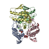
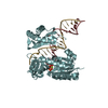
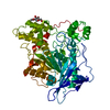
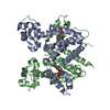
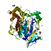
| Title | BASIC PHOSPHOLIPASE A2 FROM AGKISTRODON HALYS PALLAS-IMPLICATIONS FOR ITS ASSOCIATION AND ANTICOAGULANT ACTIVITIES BY X-RAY CRYSTALLOGRAPHY | ||||||
 Components Components | PROTEIN (PHOSPHOLIPASE A2) | ||||||
 Keywords Keywords |  HYDROLASE / BASIC PHOSPHOLIPASE A2 / AGKISTRODON HALYS PALLAS / DIMER / ANTICOAGULANT ACTIVITY HYDROLASE / BASIC PHOSPHOLIPASE A2 / AGKISTRODON HALYS PALLAS / DIMER / ANTICOAGULANT ACTIVITY | ||||||
| Function / homology |  Function and homology informationphospholipase A2 / phospholipase A2 activity / arachidonic acid secretion / phospholipid metabolic process / lipid catabolic process / toxin activity / calcium ion binding / extracellular region Function and homology informationphospholipase A2 / phospholipase A2 activity / arachidonic acid secretion / phospholipid metabolic process / lipid catabolic process / toxin activity / calcium ion binding / extracellular regionSimilarity search - Function | ||||||
| Biological species |  Gloydius halys (Halys viper) Gloydius halys (Halys viper) | ||||||
| Method | X-RAY DIFFRACTION / MOLECULAR REPLACEMENT / Resolution: 2.6 Å | ||||||
 Authors Authors | Zhao, K.H. / Lin, Z.J. | ||||||
 Citation Citation | Journal: Toxicon / Year: 2000 Title: Structure of basic phospholipase A2 from Agkistrodon halys Pallas: implications for its association, hemolytic and anticoagulant activities. Authors: Zhao, K. / Zhou, Y. / Lin, Z. #1: Journal: SCI.CHINA, SER.C: LIFE SCI. / Year: 1999Title: Refined Structure of Basic Phospholipase A2 from the Venom of Agkistrodon Halys Pallas in Orthorhombic Crystal Form I at 0.25Nm Resolution (in: C) Authors: Zhao, K.H. / Song, S.Y. / Lin, Z.J. / Zhou, Y.C. | ||||||
| History |
|
- Structure visualization
| Structure viewer | Molecule: MolmilJmol/JSmol |
|---|
- Downloads & links
-Download
| PDBx/mmCIF format | 1b4w.cif.gz | 110.5 KB | Display | PDBx/mmCIF format |
|---|---|---|---|---|
| PDB format | pdb1b4w.ent.gz | 85.8 KB | Display | PDB format |
| PDBx/mmJSON format | 1b4w.json.gz | Tree view | PDBx/mmJSON format | |
| Others |  Other downloads Other downloads |
-Validation report
| Arichive directory | https://data.pdbj.org/pub/pdb/validation_reports/b4/1b4wftp://data.pdbj.org/pub/pdb/validation_reports/b4/1b4w | HTTPS FTP |
|---|
-Related structure data
| Related structure data |  1pp2S S: Starting model for refinement |
|---|---|
| Similar structure data |


-Links
 PDBj
PDBj

- Assembly
| Deposited unit | 
| ||||||||||||||||||||||||||||
|---|---|---|---|---|---|---|---|---|---|---|---|---|---|---|---|---|---|---|---|---|---|---|---|---|---|---|---|---|---|
| 1 |
| ||||||||||||||||||||||||||||
| Unit cell |
| ||||||||||||||||||||||||||||
| Noncrystallographic symmetry (NCS) | NCS oper:
|
-Components
| #1: Protein | Mass: 13923.168 Da / Num. of mol.: 4 / Source method: isolated from a natural source / Source: (natural) Gloydius halys (Halys viper) / Cellular location: EXTRACELLULARGlossary of biology / Secretion: YES / References: UniProt: O42187, phospholipase A2#2: Sugar | Octyl glucoside  Type: D-saccharide / Mass: 292.369 Da / Num. of mol.: 2 / Source method: obtained synthetically / Formula: C14H28O6 / Comment: detergent*YM Type: D-saccharide / Mass: 292.369 Da / Num. of mol.: 2 / Source method: obtained synthetically / Formula: C14H28O6 / Comment: detergent*YM#3: Water | ChemComp-HOH / | Water Mass: 18.015 Da / Num. of mol.: 108 / Source method: isolated from a natural source / Formula: H2O Mass: 18.015 Da / Num. of mol.: 108 / Source method: isolated from a natural source / Formula: H2O |
|---|
-Experimental details
-Experiment
| Experiment | Method: X-RAY DIFFRACTION / Number of used crystals: 1 |
|---|
- Sample preparation
| Crystal | Density Matthews: 2.45 Å3/Da / Density % sol: 50 % Description: THE STRUCTURE OF BASIC PLA2 FROM AGKISTRODON HALYS PALLAS IN C2 CRYSTAL FORM WAS DETERMINED BY MOLECULAR REPLACEMENT METHOD, USING A CHAIN STRUCTURE OF THE SAME ENZYME IN P212121 CRYSTAL ...Description: THE STRUCTURE OF BASIC PLA2 FROM AGKISTRODON HALYS PALLAS IN C2 CRYSTAL FORM WAS DETERMINED BY MOLECULAR REPLACEMENT METHOD, USING A CHAIN STRUCTURE OF THE SAME ENZYME IN P212121 CRYSTAL FORM (PDB CODE 1JIA) AS SEARCH MODEL. | ||||||||||||||||||||||||||||||||||||||||||
|---|---|---|---|---|---|---|---|---|---|---|---|---|---|---|---|---|---|---|---|---|---|---|---|---|---|---|---|---|---|---|---|---|---|---|---|---|---|---|---|---|---|---|---|
| Crystal grow | pH: 8.5 Details: THE PROTEIN SOLUTIONS CONTAINED 0.1M LI2SO4, 9% PEG 4K AND 0.3% N-OCTYL BETA- D-GLUCOPYRANOSIDE IN 0.01M TRIS-HCL BUFFER(PH 8.5) AND AN ENZYME CONCENTRATION OF 8MG/ML; THE SOLUTION IN ...Details: THE PROTEIN SOLUTIONS CONTAINED 0.1M LI2SO4, 9% PEG 4K AND 0.3% N-OCTYL BETA- D-GLUCOPYRANOSIDE IN 0.01M TRIS-HCL BUFFER(PH 8.5) AND AN ENZYME CONCENTRATION OF 8MG/ML; THE SOLUTION IN RESERVOIR CONTAINED 18% PEG 4K IN SAME BUFFER, ROOM TEMPERATURE OF 17DEG.C. | ||||||||||||||||||||||||||||||||||||||||||
| Crystal grow | *PLUS Method: vapor diffusion | ||||||||||||||||||||||||||||||||||||||||||
| Components of the solutions | *PLUS
|
-Data collection
| Diffraction | Mean temperature: 280 K |
|---|---|
| Diffraction source | Source: ROTATING ANODE / Type: RIGAKU RU200 / Wavelength: 1.5418 |
| Detector | Type: SIEMENS / Detector: AREA DETECTOR / Date: Aug 15, 1997 |
| Radiation | Monochromator: NICKEL / Protocol: SINGLE WAVELENGTH / Monochromatic (M) / Laue (L): M / Scattering type: x-ray |
| Radiation wavelength | Wavelength: 1.5418 Å / Relative weight: 1 |
| Reflection | Resolution: 2.6→50 Å / Num. obs: 16462 / % possible obs: 97.3 % / Observed criterion σ(I): 0 / Redundancy: 2.6 % / Biso Wilson estimate: 38 Å2 / Rmerge(I) obs: 0.102 / Rsym value: 0.087 / Net I/σ(I): 10.4 |
| Reflection shell | Resolution: 2.6→2.8 Å / Redundancy: 2.7 % / Rmerge(I) obs: 0.381 / Mean I/σ(I) obs: 2.6 / Rsym value: 0.368 / % possible all: 96.3 |
| Reflection | *PLUS Lowest resolution: 50 Å / Num. measured all: 42427 |
| Reflection shell | *PLUS % possible obs: 93.1 % |
- Processing
| Software |
| ||||||||||||||||||||||||||||||||||||||||||||||||||||||||||||
|---|---|---|---|---|---|---|---|---|---|---|---|---|---|---|---|---|---|---|---|---|---|---|---|---|---|---|---|---|---|---|---|---|---|---|---|---|---|---|---|---|---|---|---|---|---|---|---|---|---|---|---|---|---|---|---|---|---|---|---|---|---|
| Refinement | Method to determine structure: MOLECULAR REPLACEMENT Starting model: PDB ENTRY 1PP2 Resolution: 2.6→8 Å / Cross valid method: THROUGHOUT / σ(F): 2
| ||||||||||||||||||||||||||||||||||||||||||||||||||||||||||||
| Displacement parameters | Biso mean: 25.9 Å2 | ||||||||||||||||||||||||||||||||||||||||||||||||||||||||||||
| Refine analyze | Luzzati coordinate error obs: 0.26 Å / Luzzati d res low obs: 6 Å | ||||||||||||||||||||||||||||||||||||||||||||||||||||||||||||
| Refinement step | Cycle: LAST / Resolution: 2.6→8 Å
| ||||||||||||||||||||||||||||||||||||||||||||||||||||||||||||
| Refine LS restraints |
| ||||||||||||||||||||||||||||||||||||||||||||||||||||||||||||
| Refine LS restraints NCS | NCS model details: RESTRAINTS | ||||||||||||||||||||||||||||||||||||||||||||||||||||||||||||
| LS refinement shell | Resolution: 2.6→2.71 Å / Total num. of bins used: 1
| ||||||||||||||||||||||||||||||||||||||||||||||||||||||||||||
| Xplor file |
| ||||||||||||||||||||||||||||||||||||||||||||||||||||||||||||
| Software | *PLUS Name: X-PLOR / Version: 3.8 / Classification: refinement | ||||||||||||||||||||||||||||||||||||||||||||||||||||||||||||
| Refinement | *PLUS σ(F): 2 / % reflection Rfree: 9.63 % | ||||||||||||||||||||||||||||||||||||||||||||||||||||||||||||
| Solvent computation | *PLUS | ||||||||||||||||||||||||||||||||||||||||||||||||||||||||||||
| Displacement parameters | *PLUS Biso mean: 25.9 Å2 | ||||||||||||||||||||||||||||||||||||||||||||||||||||||||||||
| Refine LS restraints | *PLUS
| ||||||||||||||||||||||||||||||||||||||||||||||||||||||||||||
| LS refinement shell | *PLUS Rfactor Rfree: 0.387 / % reflection Rfree: 10.6 % / Rfactor Rwork: 0.314 |
