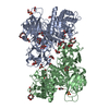
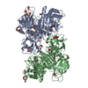
[English] 日本語
Yorodumi
- PDB-1b47: STRUCTURE OF THE N-TERMINAL DOMAIN OF CBL IN COMPLEX WITH ITS BIN... -

+
Open data


ID or keywords:

Loading...

-
Basic information

Entry
Database: PDB / ID: 1b47
TitleSTRUCTURE OF THE N-TERMINAL DOMAIN OF CBL IN COMPLEX WITH ITS BINDING SITE IN ZAP-70
ComponentsCBL
KeywordsSIGNAL TRANSDUCTION / CBL / SH2 / PROTO-ONCOGENE
Function / homology
Function and homology information


regulation of platelet-derived growth factor receptor-alpha signaling pathway / ubiquitin-dependent endocytosis / regulation of Rap protein signal transduction / entry of bacterium into host cell / flotillin complex / phosphatidylinositol 3-kinase regulatory subunit binding / positive regulation of epidermal growth factor receptor signaling pathway / Regulation of KIT signaling / mast cell degranulation / Interleukin-6 signaling ...regulation of platelet-derived growth factor receptor-alpha signaling pathway / ubiquitin-dependent endocytosis / regulation of Rap protein signal transduction / entry of bacterium into host cell / flotillin complex / phosphatidylinositol 3-kinase regulatory subunit binding / positive regulation of epidermal growth factor receptor signaling pathway / Regulation of KIT signaling / mast cell degranulation / Interleukin-6 signaling / response to testosterone / cellular response to platelet-derived growth factor stimulus / negative regulation of epidermal growth factor receptor signaling pathway / response to starvation / TGF-beta receptor signaling activates SMADs / protein monoubiquitination / protein autoubiquitination / FLT3 signaling by CBL mutants / PTK6 Regulates RTKs and Their Effectors AKT1 and DOK1 / Negative regulation of FLT3 / phosphotyrosine residue binding / ephrin receptor binding / InlB-mediated entry of Listeria monocytogenes into host cell / cellular response to nerve growth factor stimulus / response to activity / Regulation of signaling by CBL / Negative regulation of FGFR3 signaling / response to gamma radiation / Negative regulation of FGFR2 signaling / Negative regulation of FGFR4 signaling / EGFR downregulation / Negative regulation of FGFR1 signaling / Spry regulation of FGF signaling / Constitutive Signaling by EGFRvIII / RING-type E3 ubiquitin transferase / Negative regulation of MET activity / cilium / receptor tyrosine kinase binding / cytokine-mediated signaling pathway / SH3 domain binding / positive regulation of receptor-mediated endocytosis / protein polyubiquitination / ubiquitin-protein transferase activity / Signaling by CSF1 (M-CSF) in myeloid cells / male gonad development / ubiquitin protein ligase activity / Cargo recognition for clathrin-mediated endocytosis / Constitutive Signaling by Ligand-Responsive EGFR Cancer Variants / Clathrin-mediated endocytosis / growth cone / cellular response to hypoxia / ubiquitin-dependent protein catabolic process / response to ethanol / positive regulation of phosphatidylinositol 3-kinase/protein kinase B signal transduction / protein ubiquitination / cadherin binding / membrane raft / focal adhesion / DNA damage response / calcium ion binding / negative regulation of apoptotic process / perinuclear region of cytoplasm / Golgi apparatus / signal transduction / plasma membrane / cytosol
Similarity search - Function
Adaptor protein Cbl, N-terminal domain / Adaptor protein Cbl, N-terminal helical / Adaptor protein Cbl, EF hand-like / Adaptor protein Cbl, SH2-like domain / Adaptor protein Cbl, PTB domain / Adaptor protein Cbl / CBL proto-oncogene N-terminal domain 1 / CBL proto-oncogene N-terminus, EF hand-like domain / CBL proto-oncogene N-terminus, SH2-like domain / Cbl-type phosphotyrosine-binding (Cbl-PTB) domain profile. ...Adaptor protein Cbl, N-terminal domain / Adaptor protein Cbl, N-terminal helical / Adaptor protein Cbl, EF hand-like / Adaptor protein Cbl, SH2-like domain / Adaptor protein Cbl, PTB domain / Adaptor protein Cbl / CBL proto-oncogene N-terminal domain 1 / CBL proto-oncogene N-terminus, EF hand-like domain / CBL proto-oncogene N-terminus, SH2-like domain / Cbl-type phosphotyrosine-binding (Cbl-PTB) domain profile. / Adaptor protein Cbl, N-terminal domain superfamily / Transcription Elongation Factor S-II; Chain A / UBA/TS-N domain / Ubiquitin associated domain / Zinc finger, C3HC4 RING-type / Zinc finger, C3HC4 type (RING finger) / Ubiquitin-associated domain / Ubiquitin-associated domain (UBA) profile. / SH2 domain / UBA-like superfamily / SHC Adaptor Protein / EF-hand / Zinc finger, RING-type, conserved site / Zinc finger RING-type signature. / Recoverin; domain 1 / Ring finger / Zinc finger RING-type profile. / Zinc finger, RING-type / SH2 domain superfamily / EF-hand domain pair / Zinc finger, RING/FYVE/PHD-type / Up-down Bundle / 2-Layer Sandwich / Orthogonal Bundle / Mainly Alpha / Alpha Beta
Similarity search - Domain/homology
E3 ubiquitin-protein ligase CBL
Similarity search - Component
Biological speciesHomo sapiens (human)
MethodX-RAY DIFFRACTION / SYNCHROTRON / SIRAS / Resolution: 2.2 Å
AuthorsMeng, W. / Sawasdikosol, S. / Burakoff, S.J. / Eck, M.J.
CitationJournal: Nature / Year: 1999
Title: Structure of the amino-terminal domain of Cbl complexed to its binding site on ZAP-70 kinase.
Authors: Meng, W. / Sawasdikosol, S. / Burakoff, S.J. / Eck, M.J.
History
DepositionJan 6, 1999Processing site: BNL
Revision 1.0Apr 27, 1999Provider: repository / Type: Initial release
Revision 1.1Mar 24, 2008Group: Version format compliance
Revision 1.2Jul 13, 2011Group: Version format compliance
Revision 1.3Feb 7, 2024Group: Data collection / Database references ...Data collection / Database references / Derived calculations / Other
Category: chem_comp_atom / chem_comp_bond ...chem_comp_atom / chem_comp_bond / database_2 / pdbx_database_status / pdbx_struct_conn_angle / struct_conn / struct_site
Item: _database_2.pdbx_DOI / _database_2.pdbx_database_accession ..._database_2.pdbx_DOI / _database_2.pdbx_database_accession / _pdbx_database_status.process_site / _pdbx_struct_conn_angle.ptnr1_auth_comp_id / _pdbx_struct_conn_angle.ptnr1_auth_seq_id / _pdbx_struct_conn_angle.ptnr1_label_atom_id / _pdbx_struct_conn_angle.ptnr1_label_comp_id / _pdbx_struct_conn_angle.ptnr1_label_seq_id / _pdbx_struct_conn_angle.ptnr3_auth_comp_id / _pdbx_struct_conn_angle.ptnr3_auth_seq_id / _pdbx_struct_conn_angle.ptnr3_label_atom_id / _pdbx_struct_conn_angle.ptnr3_label_comp_id / _pdbx_struct_conn_angle.ptnr3_label_seq_id / _pdbx_struct_conn_angle.value / _struct_conn.pdbx_dist_value / _struct_conn.ptnr1_auth_asym_id / _struct_conn.ptnr1_auth_comp_id / _struct_conn.ptnr1_auth_seq_id / _struct_conn.ptnr1_label_asym_id / _struct_conn.ptnr1_label_atom_id / _struct_conn.ptnr1_label_comp_id / _struct_conn.ptnr1_label_seq_id / _struct_conn.ptnr2_auth_asym_id / _struct_conn.ptnr2_auth_comp_id / _struct_conn.ptnr2_auth_seq_id / _struct_conn.ptnr2_label_asym_id / _struct_conn.ptnr2_label_atom_id / _struct_conn.ptnr2_label_comp_id / _struct_conn.ptnr2_label_seq_id / _struct_site.pdbx_auth_asym_id / _struct_site.pdbx_auth_comp_id / _struct_site.pdbx_auth_seq_id

-
Structure visualization

Structure viewerMolecule:
MolmilJmol/JSmol

Downloads & links

-
Assembly

Deposited unit
A: CBL
B: CBL
C: CBL
hetero molecules


Theoretical massNumber of molelcules
Total (without water)106,2396
Polymers106,1193
Non-polymers1203
Water11,998666
1


  • Idetical with deposited unit
  • defined by author
TypeNameSymmetry operationNumber
identity operation1_555x,y,z1
Unit cell
Length a, b, c (Å)159.960, 105.480, 84.920
Angle α, β, γ (deg.)90.00, 92.06, 90.00
Int Tables number5
Space group name H-MC121
Noncrystallographic symmetry (NCS)NCS oper:
IDCodeMatrixVector
1given(-0.399672, 0.916202, -0.028918), (-0.914736, -0.396594, 0.077273), (0.059329, 0.057336, 0.996591)29.8495, 35.2372, -30.3331
2given(-0.746434, -0.66218, 0.065977), (0.664788, -0.737557, 0.118599), (-0.029872, 0.132387, 0.990748)51.2264, -11.0673, -56.081

-
Components

#1: Protein CBL / PROTO-ONCOGENE C-CBL


Mass: 35372.910 Da / Num. of mol.: 3 / Fragment: N-TERMINAL DOMAIN
Source method: isolated from a genetically manipulated source
Source: (gene. exp.) Homo sapiens (human) / Cellular location: CYTOPLASM / Plasmid: PGEX 4T-3 / Production host: Escherichia coli (E. coli) / Strain (production host): DH5 ALPHA / References: UniProt: P22681
#2: Chemical ChemComp-CA / CALCIUM ION


Mass: 40.078 Da / Num. of mol.: 3 / Source method: obtained synthetically / Formula: Ca
#3: Water ChemComp-HOH / water / Water


Mass: 18.015 Da / Num. of mol.: 666 / Source method: isolated from a natural source / Formula: H2O

-
Experimental details

-
Experiment

ExperimentMethod: X-RAY DIFFRACTION / Number of used crystals: 20

-
Sample preparation

CrystalDensity Matthews: 3.15 Å3/Da / Density % sol: 60 %
Crystal growpH: 5.6 / Details: pH 5.6
Crystal
*PLUS
Crystal grow
*PLUS
Temperature: 22 ℃ / Method: vapor diffusion, hanging drop
Components of the solutions
*PLUS
IDConc.Common nameCrystal-IDSol-ID
120 %PEG80001reservoir
20.2 Mcalcium acetate1reservoir
32 mMdithiothreitol1reservoir
40.1 Msodium cacodylate1reservoir

-
Data collection

DiffractionMean temperature: 100 K
Diffraction sourceSource: SYNCHROTRON / Site: CHESS / Beamline: F1 / Wavelength: 0.918
DetectorType: ADSC / Detector: CCD / Date: Dec 1, 1997
RadiationMonochromatic (M) / Laue (L): M / Scattering type: x-ray
Radiation wavelengthWavelength: 0.918 Å / Relative weight: 1
ReflectionResolution: 2.2→50 Å / Num. obs: 70918 / % possible obs: 99.3 % / Redundancy: 2.5 % / Biso Wilson estimate: 45.12 Å2 / Rsym value: 0.067 / Net I/σ(I): 3.9
Reflection shellResolution: 2.2→2.32 Å / Redundancy: 2.5 % / Mean I/σ(I) obs: 1.1 / Rsym value: 0.391 / % possible all: 99.4
Reflection
*PLUS
Num. measured all: 178845 / Rmerge(I) obs: 0.067

-
Processing

Software
NameVersionClassification
HEAVYmodel building
PATSOLmodel building
CNS0.4refinement
MOSFLMdata reduction
CCP4data scaling
HEAVYphasing
PATSOLphasing
RefinementMethod to determine structure: SIRAS / Resolution: 2.2→50 Å / Data cutoff high rms absF: 10000 / Isotropic thermal model: RESTRAINED / Cross valid method: THROUGHOUT / σ(F): 0
RfactorNum. reflection% reflectionSelection details
Rfree0.266 3595 5 %RANDOM
Rwork0.218 ---
obs0.218 70918 99.3 %-
Displacement parametersBiso mean: 47.6 Å2
Baniso -1Baniso -2Baniso -3
1--3.489 Å20 Å20.066 Å2
2---4.825 Å20 Å2
3---8.314 Å2
Refine analyze
FreeObs
Luzzati coordinate error0.37 Å0.28 Å
Luzzati d res low-5 Å
Luzzati sigma a0.36 Å0.24 Å
Refinement stepCycle: LAST / Resolution: 2.2→50 Å
ProteinNucleic acidLigandSolventTotal
Num. atoms8092 0 3 666 8761
Refine LS restraints
Refine-IDTypeDev idealDev ideal target
X-RAY DIFFRACTIONc_bond_d0.018
X-RAY DIFFRACTIONc_bond_d_na
X-RAY DIFFRACTIONc_bond_d_prot
X-RAY DIFFRACTIONc_angle_d
X-RAY DIFFRACTIONc_angle_d_na
X-RAY DIFFRACTIONc_angle_d_prot
X-RAY DIFFRACTIONc_angle_deg2.61
X-RAY DIFFRACTIONc_angle_deg_na
X-RAY DIFFRACTIONc_angle_deg_prot
X-RAY DIFFRACTIONc_dihedral_angle_d24.53
X-RAY DIFFRACTIONc_dihedral_angle_d_na
X-RAY DIFFRACTIONc_dihedral_angle_d_prot
X-RAY DIFFRACTIONc_improper_angle_d4.22
X-RAY DIFFRACTIONc_improper_angle_d_na
X-RAY DIFFRACTIONc_improper_angle_d_prot
X-RAY DIFFRACTIONc_mcbond_it1.1871.5
X-RAY DIFFRACTIONc_mcangle_it1.9132
X-RAY DIFFRACTIONc_scbond_it1.9272
X-RAY DIFFRACTIONc_scangle_it2.8452.5
LS refinement shellResolution: 2.2→2.28 Å / Total num. of bins used: 10
RfactorNum. reflection% reflection
Rfree0.36 374 5 %
Rwork0.28 6707 -
obs--99 %
Xplor file
Refine-IDSerial noParam file
X-RAY DIFFRACTION1CNS_TOPPAR:PROTEIN_REP.PA
X-RAY DIFFRACTION2CNS_TOPPAR:WATER_REP.PARA
Software
*PLUS
Name: CNS / Version: 0.4 / Classification: refinement
Refine LS restraints
*PLUS
Refine-IDTypeDev ideal
X-RAY DIFFRACTIONc_dihedral_angle_d
X-RAY DIFFRACTIONc_dihedral_angle_deg24.53
X-RAY DIFFRACTIONc_improper_angle_d
X-RAY DIFFRACTIONc_improper_angle_deg4.22

+
About Yorodumi

-
News

-
Feb 9, 2022. New format data for meta-information of EMDB entries

New format data for meta-information of EMDB entries

  • Version 3 of the EMDB header file is now the official format.
  • The previous official version 1.9 will be removed from the archive.

Related info.:EMDB header

External links:wwPDB to switch to version 3 of the EMDB data model

-
Aug 12, 2020. Covid-19 info

Covid-19 info

URL: https://pdbj.org/emnavi/covid19.php

New page: Covid-19 featured information page in EM Navigator.

Related info.:Covid-19 info / Mar 5, 2020. Novel coronavirus structure data

+
Mar 5, 2020. Novel coronavirus structure data

Novel coronavirus structure data

Related info.:Yorodumi Speices / Aug 12, 2020. Covid-19 info

External links:COVID-19 featured content - PDBj / Molecule of the Month (242):Coronavirus Proteases

+
Jan 31, 2019. EMDB accession codes are about to change! (news from PDBe EMDB page)

EMDB accession codes are about to change! (news from PDBe EMDB page)

  • The allocation of 4 digits for EMDB accession codes will soon come to an end. Whilst these codes will remain in use, new EMDB accession codes will include an additional digit and will expand incrementally as the available range of codes is exhausted. The current 4-digit format prefixed with “EMD-” (i.e. EMD-XXXX) will advance to a 5-digit format (i.e. EMD-XXXXX), and so on. It is currently estimated that the 4-digit codes will be depleted around Spring 2019, at which point the 5-digit format will come into force.
  • The EM Navigator/Yorodumi systems omit the EMD- prefix.

Related info.:Q: What is EMD? / ID/Accession-code notation in Yorodumi/EM Navigator

External links:EMDB Accession Codes are Changing Soon! / Contact to PDBj

+
Jul 12, 2017. Major update of PDB

Major update of PDB

  • wwPDB released updated PDB data conforming to the new PDBx/mmCIF dictionary.
  • This is a major update changing the version number from 4 to 5, and with Remediation, in which all the entries are updated.
  • In this update, many items about electron microscopy experimental information are reorganized (e.g. em_software).
  • Now, EM Navigator and Yorodumi are based on the updated data.

External links:wwPDB Remediation / Enriched Model Files Conforming to OneDep Data Standards Now Available in the PDB FTP Archive

-
Yorodumi

Thousand views of thousand structures

  • Yorodumi is a browser for structure data from EMDB, PDB, SASBDB, etc.
  • This page is also the successor to EM Navigator detail page, and also detail information page/front-end page for Omokage search.
  • The word "yorodu" (or yorozu) is an old Japanese word meaning "ten thousand". "mi" (miru) is to see.

Related info.:EMDB / PDB / SASBDB / Comparison of 3 databanks / Yorodumi Search / Aug 31, 2016. New EM Navigator & Yorodumi / Yorodumi Papers / Jmol/JSmol / Function and homology information / Changes in new EM Navigator and Yorodumi

Read more