 Movie
Movie Controller

[English] 日本語
 Yorodumi
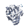
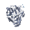
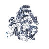
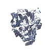
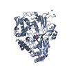
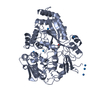
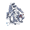
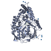
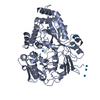
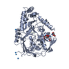
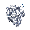
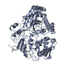
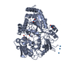
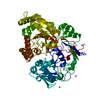
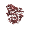
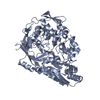
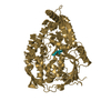
- PDB-1b3h: OLIGO-PEPTIDE BINDING PROTEIN COMPLEXED WITH LYSYL-CYCLOHEXYLALAN... -
+ Open data

- Basic information
| Entry | Database: PDB / ID: 1b3h | ||||||
|---|---|---|---|---|---|---|---|
| Title | OLIGO-PEPTIDE BINDING PROTEIN COMPLEXED WITH LYSYL-CYCLOHEXYLALANYL-LYSINE | ||||||
 Components Components |
| ||||||
 Keywords Keywords | PEPTIDE BINDING PROTEIN / PERIPLASMIC PEPTIDE BINDING PROTEIN | ||||||
| Function / homology |  Function and homology information Function and homology informationpeptide transport / peptide transmembrane transporter activity / ATP-binding cassette (ABC) transporter complex /  protein transport / outer membrane-bounded periplasmic space protein transport / outer membrane-bounded periplasmic spaceSimilarity search - Function | ||||||
| Biological species |  Salmonella typhimurium (bacteria) Salmonella typhimurium (bacteria) | ||||||
| Method | X-RAY DIFFRACTION / MOLECULAR REPLACEMENT / Resolution: 2 Å | ||||||
 Authors Authors | Davies, T.G. / Tame, J.R.H. | ||||||
 Citation Citation | Journal: Protein Sci. / Year: 1999 Title: Relating structure to thermodynamics: the crystal structures and binding affinity of eight OppA-peptide complexes. Authors: Davies, T.G. / Hubbard, R.E. / Tame, J.R. #1: Journal: Structure / Year: 1995Title: The Crystal Structures of the Oligopeptide-Binding Protein Oppa Complexed with Tripeptide and Tetrapeptide Ligands Authors: Tame, J.R. / Dodson, E.J. / Murshudov, G. / Higgins, C.F. / Wilkinson, A.J. | ||||||
| History |
|
- Structure visualization
| Structure viewer | Molecule: MolmilJmol/JSmol |
|---|
- Downloads & links
-Download
| PDBx/mmCIF format | 1b3h.cif.gz | 120.5 KB | Display | PDBx/mmCIF format |
|---|---|---|---|---|
| PDB format | pdb1b3h.ent.gz | 92.8 KB | Display | PDB format |
| PDBx/mmJSON format | 1b3h.json.gz | Tree view | PDBx/mmJSON format | |
| Others |  Other downloads Other downloads |
-Validation report
| Arichive directory | https://data.pdbj.org/pub/pdb/validation_reports/b3/1b3hftp://data.pdbj.org/pub/pdb/validation_reports/b3/1b3h | HTTPS FTP |
|---|
-Related structure data
| Related structure data | 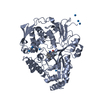 1b0hC  1b1hC  1b2hC  1b4hC  1b5hC 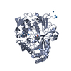 1b6hC 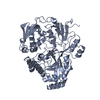 1b7hC C: citing same article ( |
|---|---|
| Similar structure data |


-Links
 PDBj
PDBj- Assembly
| Deposited unit | 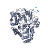
| ||||||||||
|---|---|---|---|---|---|---|---|---|---|---|---|
| 1 |
| ||||||||||
| Unit cell |
|
-Components
| #1: Protein | Mass: 58878.984 Da / Num. of mol.: 1 Source method: isolated from a genetically manipulated source Source: (gene. exp.) Salmonella typhimurium (bacteria) / Production host: Escherichia coli (E. coli) / References: UniProt: P06202 | ||||
|---|---|---|---|---|---|
| #2: Protein/peptide | Mass: 429.597 Da / Num. of mol.: 1 / Source method: obtained synthetically | ||||
| #3: Chemical | ChemComp-U1 /   Mass: 238.029 Da / Num. of mol.: 8 / Source method: obtained synthetically / Formula: U Mass: 238.029 Da / Num. of mol.: 8 / Source method: obtained synthetically / Formula: U#4: Water | ChemComp-HOH / | Water Mass: 18.015 Da / Num. of mol.: 249 / Source method: isolated from a natural source / Formula: H2O Mass: 18.015 Da / Num. of mol.: 249 / Source method: isolated from a natural source / Formula: H2ONonpolymer details | THE SOLVENT STRUCTURE AROUND THE URANIUM IONS IS TENTATIVE | |
-Experimental details
-Experiment
| Experiment | Method: X-RAY DIFFRACTION / Number of used crystals: 1 |
|---|
- Sample preparation
| Crystal | Density Matthews: 2.45 Å3/Da / Density % sol: 49.81 % | |||||||||||||||||||||||||
|---|---|---|---|---|---|---|---|---|---|---|---|---|---|---|---|---|---|---|---|---|---|---|---|---|---|---|
| Crystal grow | pH: 5.5 / Details: pH 5.5 | |||||||||||||||||||||||||
| Crystal grow | *PLUS Method: vapor diffusion, hanging drop | |||||||||||||||||||||||||
| Components of the solutions | *PLUS
|
-Data collection
| Diffraction | Mean temperature: 120 K |
|---|---|
| Diffraction source | Source: ROTATING ANODE / Wavelength: 1.5418 |
| Radiation | Protocol: SINGLE WAVELENGTH / Monochromatic (M) / Laue (L): M / Scattering type: x-ray |
| Radiation wavelength | Wavelength: 1.5418 Å / Relative weight: 1 |
| Reflection | Resolution: 2→20 Å / Num. obs: 38674 / % possible obs: 98 % / Redundancy: 6 % / Rmerge(I) obs: 0.135 / Net I/σ(I): 3.7 |
| Reflection | *PLUS % possible obs: 98.3 % / Num. measured all: 231066 |
| Reflection shell | *PLUS % possible obs: 91.3 % / Rmerge(I) obs: 0.32 / Mean I/σ(I) obs: 2 |
- Processing
| Software |
| ||||||||||||||||||||||||||||||||||||||||||||||||||||||||||||||||||||||||||||||||||||
|---|---|---|---|---|---|---|---|---|---|---|---|---|---|---|---|---|---|---|---|---|---|---|---|---|---|---|---|---|---|---|---|---|---|---|---|---|---|---|---|---|---|---|---|---|---|---|---|---|---|---|---|---|---|---|---|---|---|---|---|---|---|---|---|---|---|---|---|---|---|---|---|---|---|---|---|---|---|---|---|---|---|---|---|---|---|
| Refinement | Method to determine structure: MOLECULAR REPLACEMENT / Resolution: 2→20 Å / Cross valid method: THROUGHOUT / σ(F): 0
| ||||||||||||||||||||||||||||||||||||||||||||||||||||||||||||||||||||||||||||||||||||
| Displacement parameters | Biso mean: 36 Å2 | ||||||||||||||||||||||||||||||||||||||||||||||||||||||||||||||||||||||||||||||||||||
| Refinement step | Cycle: LAST / Resolution: 2→20 Å
| ||||||||||||||||||||||||||||||||||||||||||||||||||||||||||||||||||||||||||||||||||||
| Refine LS restraints |
| ||||||||||||||||||||||||||||||||||||||||||||||||||||||||||||||||||||||||||||||||||||
| Software | *PLUS Name: REFMAC / Classification: refinement | ||||||||||||||||||||||||||||||||||||||||||||||||||||||||||||||||||||||||||||||||||||
| Refinement | *PLUS Rfactor Rfree: 0.239 / Rfactor Rwork: 0.191 | ||||||||||||||||||||||||||||||||||||||||||||||||||||||||||||||||||||||||||||||||||||
| Solvent computation | *PLUS | ||||||||||||||||||||||||||||||||||||||||||||||||||||||||||||||||||||||||||||||||||||
| Displacement parameters | *PLUS |
