 Movie
Movie Controller


+ Open data

- Basic information
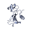
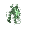
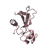
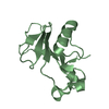
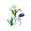
| Entry | Database: PDB / ID: 1b21 | ||||||
|---|---|---|---|---|---|---|---|
| Title | DELETION OF A BURIED SALT BRIDGE IN BARNASE | ||||||
 Components Components | PROTEIN (BARNASE) | ||||||
 Keywords Keywords |  HYDROLASE / MICROBIAL RIBONUCLEASE / ALPHA/BETA PROTEIN HYDROLASE / MICROBIAL RIBONUCLEASE / ALPHA/BETA PROTEIN | ||||||
| Function / homology |  Function and homology informationHydrolases; Acting on ester bonds; Endoribonucleases producing 3'-phosphomonoesters / RNA endonuclease activity / RNA binding / extracellular region Function and homology informationHydrolases; Acting on ester bonds; Endoribonucleases producing 3'-phosphomonoesters / RNA endonuclease activity / RNA binding / extracellular regionSimilarity search - Function | ||||||
| Biological species |  Bacillus amyloliquefaciens (bacteria) Bacillus amyloliquefaciens (bacteria) | ||||||
| Method | X-RAY DIFFRACTION / OTHER / Resolution: 2 Å | ||||||
 Authors Authors | Vaughan, C.K. / Harryson, P. / Buckle, A.M. / Oliveberg, M. / Fersht, A.R. | ||||||
 Citation Citation | Journal: Acta Crystallogr.,Sect.D / Year: 2002 Title: A structural double-mutant cycle: estimating the strength of a buried salt bridge in barnase. Authors: Vaughan, C.K. / Harryson, P. / Buckle, A.M. / Fersht, A.R. #1: Journal: Nature / Year: 1982Title: Molecular Structure of a New Family of Ribonucleases Authors: Mauguen, Y. / Hartley, R.W. / Dodson, E.J. / Dodson, G.G. / Bricogne, G. / Chothia, C. / Jack, A. | ||||||
| History |
|
- Structure visualization
| Structure viewer | Molecule: MolmilJmol/JSmol |
|---|
- Downloads & links
-Download
| PDBx/mmCIF format | 1b21.cif.gz | 83.5 KB | Display | PDBx/mmCIF format |
|---|---|---|---|---|
| PDB format | pdb1b21.ent.gz | 63.2 KB | Display | PDB format |
| PDBx/mmJSON format | 1b21.json.gz | Tree view | PDBx/mmJSON format | |
| Others |  Other downloads Other downloads |
-Validation report
| Arichive directory | https://data.pdbj.org/pub/pdb/validation_reports/b2/1b21ftp://data.pdbj.org/pub/pdb/validation_reports/b2/1b21 | HTTPS FTP |
|---|
-Related structure data


-Links
 PDBj
PDBj- Assembly
| Deposited unit | 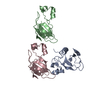
| ||||||||||||
|---|---|---|---|---|---|---|---|---|---|---|---|---|---|
| 1 | 
| ||||||||||||
| 2 | 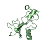
| ||||||||||||
| 3 | 
| ||||||||||||
| Unit cell |
| ||||||||||||
| Noncrystallographic symmetry (NCS) | NCS oper:
|
-Components
| #1: Protein | Mass: 12327.620 Da / Num. of mol.: 3 / Mutation: R69S, D93N Source method: isolated from a genetically manipulated source Source: (gene. exp.) Bacillus amyloliquefaciens (bacteria) / Cellular location: EXTRACELLULARGlossary of biology / Plasmid: PMT410 / Production host: Escherichia coli (E. coli) / References: UniProt: P00648, EC: 3.1.27.3#2: Chemical | ChemComp-ZN / |   Mass: 65.409 Da / Num. of mol.: 1 / Source method: obtained synthetically / Formula: Zn Mass: 65.409 Da / Num. of mol.: 1 / Source method: obtained synthetically / Formula: Zn#3: Water | ChemComp-HOH / | Water Mass: 18.015 Da / Num. of mol.: 339 / Source method: isolated from a natural source / Formula: H2O Mass: 18.015 Da / Num. of mol.: 339 / Source method: isolated from a natural source / Formula: H2O |
|---|
-Experimental details
-Experiment
| Experiment | Method: X-RAY DIFFRACTION / Number of used crystals: 1 |
|---|
- Sample preparation
| Crystal | Density Matthews: 2.11 Å3/Da / Density % sol: 42 % Description: THE STRUCTURE WAS SOLVED BY RIGID BODY REFINEMENT USING WILD-TYPE BARNASE AS THE STARTING MODEL | ||||||||||||||||||||||||||||||||||||||||||||||||
|---|---|---|---|---|---|---|---|---|---|---|---|---|---|---|---|---|---|---|---|---|---|---|---|---|---|---|---|---|---|---|---|---|---|---|---|---|---|---|---|---|---|---|---|---|---|---|---|---|---|
| Crystal grow | pH: 7.5 Details: DROP: 8-12 MG/ML PROTEIN 6 MM ZNSO4 0.6 M (NH4)2SO4 WELL: 2.58-2.73 M AMMONIUM PHOSPHATE BUFFER, PH 7.5 1-2 MM ZNSO4 0.15-0.30 M (NH4)2SO4 5-10 MM NH4OH | ||||||||||||||||||||||||||||||||||||||||||||||||
| Components of the solutions |
| ||||||||||||||||||||||||||||||||||||||||||||||||
| Crystal grow | *PLUS Temperature: 288 K / Method: vapor diffusion, hanging drop | ||||||||||||||||||||||||||||||||||||||||||||||||
| Components of the solutions | *PLUS
|
-Data collection
| Diffraction | Mean temperature: 100 K |
|---|---|
| Diffraction source | Source: ROTATING ANODE / Type: ELLIOTT GX-13 / Wavelength: 1.5418 |
| Detector | Type: MARRESEARCH / Detector: IMAGE PLATE / Date: May 1, 1997 / Details: SUPER DOUBLE MIRRORS |
| Radiation | Protocol: SINGLE WAVELENGTH / Monochromatic (M) / Laue (L): M / Scattering type: x-ray |
| Radiation wavelength | Wavelength: 1.5418 Å / Relative weight: 1 |
| Reflection | Resolution: 2.01→21.32 Å / Num. obs: 18526 / % possible obs: 91.4 % / Redundancy: 1.8 % / Biso Wilson estimate: 12.67 Å2 / Rmerge(I) obs: 0.067 / Net I/σ(I): 10.06 |
| Reflection shell | Resolution: 2.01→2.12 Å / Redundancy: 1.6 % / Rmerge(I) obs: 0.144 / Mean I/σ(I) obs: 5.1 / % possible all: 59.3 |
| Reflection | *PLUS Highest resolution: 2 Å / Rmerge(I) obs: 0.067 |
| Reflection shell | *PLUS % possible obs: 59.3 % / Rmerge(I) obs: 0.144 |
- Processing
| Software |
| ||||||||||||||||||||||||||||||||||||||||||||||||||||||||||||||||||||||||||||||||||||
|---|---|---|---|---|---|---|---|---|---|---|---|---|---|---|---|---|---|---|---|---|---|---|---|---|---|---|---|---|---|---|---|---|---|---|---|---|---|---|---|---|---|---|---|---|---|---|---|---|---|---|---|---|---|---|---|---|---|---|---|---|---|---|---|---|---|---|---|---|---|---|---|---|---|---|---|---|---|---|---|---|---|---|---|---|---|
| Refinement | Method to determine structure: OTHER / Resolution: 2→19 Å / SU B: 5.81 / Cross valid method: THROUGHOUT / σ(F): 0
| ||||||||||||||||||||||||||||||||||||||||||||||||||||||||||||||||||||||||||||||||||||
| Displacement parameters | Biso mean: 19.3 Å2 | ||||||||||||||||||||||||||||||||||||||||||||||||||||||||||||||||||||||||||||||||||||
| Refinement step | Cycle: LAST / Resolution: 2→19 Å
| ||||||||||||||||||||||||||||||||||||||||||||||||||||||||||||||||||||||||||||||||||||
| Refine LS restraints |
| ||||||||||||||||||||||||||||||||||||||||||||||||||||||||||||||||||||||||||||||||||||
| Software | *PLUS Name: REFMAC / Classification: refinement | ||||||||||||||||||||||||||||||||||||||||||||||||||||||||||||||||||||||||||||||||||||
| Refinement | *PLUS Highest resolution: 2 Å / σ(F): 0 / % reflection Rfree: 5 % / Rfactor obs: 0.188 / Rfactor Rfree: 0.268 / Rfactor Rwork: 0.188 | ||||||||||||||||||||||||||||||||||||||||||||||||||||||||||||||||||||||||||||||||||||
| Solvent computation | *PLUS | ||||||||||||||||||||||||||||||||||||||||||||||||||||||||||||||||||||||||||||||||||||
| Displacement parameters | *PLUS Biso mean: 19.3 Å2 |
