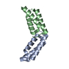
Entry Database : PDB / ID : 1b0xTitle THE CRYSTAL STRUCTURE OF AN EPH RECEPTOR SAM DOMAIN REVEALS A MECHANISM FOR MODULAR DIMERIZATION. PROTEIN (EPHA4 RECEPTOR TYROSINE KINASE) Keywords / / / Function / homology Function Domain/homology Component
/ / / / / / / / / / / / / / / / / / / / / / / / / / / / / / / / / / / / / / / / / / / / / / / / / / / / / / / / / / / / / / / / / / / / / / / / / / / / / / / / / / / / / / / / / / / / / / / / / / / / / / / / / / / / / / / / / / / / / / / / / / / / / / / / / / / / / / / / / / / Biological species Mus musculus (house mouse)Method / / Resolution : 2 Å Authors Stapleton, D. / Balan, I. / Pawson, T. / Sicheri, F. Journal : Nat.Struct.Biol. / Year : 1999Title : The crystal structure of an Eph receptor SAM domain reveals a mechanism for modular dimerization.Authors : Stapleton, D. / Balan, I. / Pawson, T. / Sicheri, F. History Deposition Nov 14, 1998 Deposition site / Processing site Revision 1.0 May 3, 1999 Provider / Type Revision 1.1 Apr 26, 2008 Group Revision 1.2 Jul 13, 2011 Group Revision 1.3 Dec 27, 2023 Group / Database referencesCategory chem_comp_atom / chem_comp_bond ... chem_comp_atom / chem_comp_bond / database_2 / struct_ref_seq_dif Item / _database_2.pdbx_database_accession / _struct_ref_seq_dif.details
Show all Show less
 Movie
Movie Controller

 Yorodumi
 Open data
 Basic information
 Components
 Keywords
 TRANSFERASE /
  Function and homology information
 Authors
 Citation
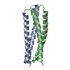
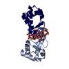
 Structure visualization
 Downloads & links
 Other downloads
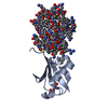


 PDBj
PDBj


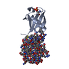
 Assembly


 Mass: 18.015 Da / Num. of mol.: 53 / Source method: isolated from a natural source / Formula: H2O
 Sample preparation
 Processing