 Movie
Movie Controller


+ Open data

- Basic information
| Entry | Database: PDB / ID: 1awr | ||||||
|---|---|---|---|---|---|---|---|
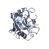
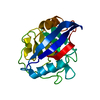
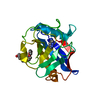
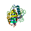
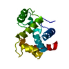
| Title | CYPA COMPLEXED WITH HAGPIA | ||||||
 Components Components |
| ||||||
 Keywords Keywords | COMPLEX (ISOMERASE/PEPTIDE) / COMPLEX (ISOMERASE-PEPTIDE) /  CYCLOPHILIN A / HIV-1 CAPSID / PSEUDO-SYMMETRY / COMPLEX (ISOMERASE-PEPTIDE) complex CYCLOPHILIN A / HIV-1 CAPSID / PSEUDO-SYMMETRY / COMPLEX (ISOMERASE-PEPTIDE) complex | ||||||
| Function / homology |  Function and homology information Function and homology informationnegative regulation of protein K48-linked ubiquitination / negative regulation of viral life cycle / regulation of apoptotic signaling pathway / cell adhesion molecule production / lipid droplet organization / heparan sulfate binding / regulation of viral genome replication / leukocyte chemotaxis / negative regulation of stress-activated MAPK cascade / endothelial cell activation ...negative regulation of protein K48-linked ubiquitination / negative regulation of viral life cycle / regulation of apoptotic signaling pathway / cell adhesion molecule production / lipid droplet organization / heparan sulfate binding / regulation of viral genome replication / leukocyte chemotaxis / negative regulation of stress-activated MAPK cascade / endothelial cell activation / virion binding / Basigin interactions / cyclosporin A binding / Minus-strand DNA synthesis / Plus-strand DNA synthesis / Uncoating of the HIV Virion / Early Phase of HIV Life Cycle / Integration of provirus / APOBEC3G mediated resistance to HIV-1 infection / Calcineurin activates NFAT / viral release from host cell / Binding and entry of HIV virion / positive regulation of viral genome replication / protein peptidyl-prolyl isomerization / negative regulation of oxidative stress-induced intrinsic apoptotic signaling pathway / positive regulation of protein dephosphorylation / Gene and protein expression by JAK-STAT signaling after Interleukin-12 stimulation / activation of protein kinase B activity / neutrophil chemotaxis / negative regulation of protein phosphorylation / peptidylprolyl isomerase / peptidyl-prolyl cis-trans isomerase activity / positive regulation of protein secretion / negative regulation of protein kinase activity / Assembly Of The HIV Virion / Budding and maturation of HIV virion / neuron differentiation / platelet activation / platelet aggregation / SARS-CoV-1 activates/modulates innate immune responses / unfolded protein binding / integrin binding / protein folding / Platelet degranulation / cellular response to oxidative stress / positive regulation of NF-kappaB transcription factor activity / secretory granule lumen / vesicle / ficolin-1-rich granule lumen / positive regulation of MAPK cascade / positive regulation of protein phosphorylation / focal adhesion / apoptotic process / Neutrophil degranulation / protein-containing complex / extracellular space / RNA binding / extracellular exosome / extracellular region / membrane / nucleus / cytosol / cytoplasmSimilarity search - Function | ||||||
| Biological species |  Homo sapiens (human) Homo sapiens (human) | ||||||
| Method | X-RAY DIFFRACTION / SYNCHROTRON / MOLECULAR REPLACEMENT / Resolution: 1.58 Å | ||||||
 Authors Authors | Vajdos, F.F. | ||||||
 Citation Citation | Journal: Protein Sci. / Year: 1997 Title: Crystal structure of cyclophilin A complexed with a binding site peptide from the HIV-1 capsid protein. Authors: Vajdos, F.F. / Yoo, S. / Houseweart, M. / Sundquist, W.I. / Hill, C.P. | ||||||
| History |
|
- Structure visualization
| Structure viewer | Molecule: MolmilJmol/JSmol |
|---|
- Downloads & links
-Download
| PDBx/mmCIF format | 1awr.cif.gz | 196.9 KB | Display | PDBx/mmCIF format |
|---|---|---|---|---|
| PDB format | pdb1awr.ent.gz | 160.9 KB | Display | PDB format |
| PDBx/mmJSON format | 1awr.json.gz | Tree view | PDBx/mmJSON format | |
| Others |  Other downloads Other downloads |
-Validation report
| Arichive directory | https://data.pdbj.org/pub/pdb/validation_reports/aw/1awrftp://data.pdbj.org/pub/pdb/validation_reports/aw/1awr | HTTPS FTP |
|---|
-Related structure data
| Related structure data |  1awqC  1awsC  1awtC  1awuC  1awvC  2cyhS S: Starting model for refinement C: citing same article ( |
|---|---|
| Similar structure data |


-Links
 PDBj
PDBj


- Assembly
| Deposited unit | 
| ||||||||||||||||||||||||
|---|---|---|---|---|---|---|---|---|---|---|---|---|---|---|---|---|---|---|---|---|---|---|---|---|---|
| 1 | 
| ||||||||||||||||||||||||
| 2 | 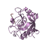
| ||||||||||||||||||||||||
| 3 | 
| ||||||||||||||||||||||||
| 4 | 
| ||||||||||||||||||||||||
| 5 | 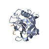
| ||||||||||||||||||||||||
| 6 | 
| ||||||||||||||||||||||||
| Unit cell |
| ||||||||||||||||||||||||
| Noncrystallographic symmetry (NCS) | NCS oper:
|
-Components
| #1: Protein | Peptidylprolyl isomerase A Mass: 17905.307 Da / Num. of mol.: 6 Source method: isolated from a genetically manipulated source Source: (gene. exp.) Homo sapiens (human) / Cellular location: CYTOPLASM / Gene: CYCLOPHILIN / Gene (production host): CYCLOPHILIN / Production host:  Escherichia coli (E. coli) / References: UniProt: P62937, peptidylprolyl isomerase Escherichia coli (E. coli) / References: UniProt: P62937, peptidylprolyl isomerase#2: Protein/peptide | Mass: 565.642 Da / Num. of mol.: 6 Source method: isolated from a genetically manipulated source #3: Water | ChemComp-HOH / | Water Mass: 18.015 Da / Num. of mol.: 175 / Source method: isolated from a natural source / Formula: H2O Mass: 18.015 Da / Num. of mol.: 175 / Source method: isolated from a natural source / Formula: H2O |
|---|
-Experimental details
-Experiment
| Experiment | Method: X-RAY DIFFRACTION / Number of used crystals: 1 |
|---|
- Sample preparation
| Crystal | Density Matthews: 2.26 Å3/Da / Density % sol: 45.58 % | ||||||||||||||||||||
|---|---|---|---|---|---|---|---|---|---|---|---|---|---|---|---|---|---|---|---|---|---|
| Crystal grow | pH: 8.4 / Details: pH 8.4 | ||||||||||||||||||||
| Crystal grow | *PLUS Temperature: 21 ℃ / Method: unknown | ||||||||||||||||||||
| Components of the solutions | *PLUS
|
-Data collection
| Diffraction | Mean temperature: 100 K |
|---|---|
| Diffraction source | Source: SYNCHROTRON / Site: NSLS  / Beamline: X12C / Wavelength: 1.1 / Beamline: X12C / Wavelength: 1.1 |
| Detector | Type: MARRESEARCH / Detector: IMAGE PLATE / Date: Oct 28, 1995 |
| Radiation | Monochromator: SI(111) / Monochromatic (M) / Laue (L): M / Scattering type: x-ray |
| Radiation wavelength | Wavelength: 1.1 Å / Relative weight: 1 |
| Reflection | Resolution: 1.58→15 Å / Num. obs: 147562 / % possible obs: 88.4 % / Observed criterion σ(I): 0 / Biso Wilson estimate: 16.4 Å2 / Rsym value: 0.088 / Net I/σ(I): 4.2 |
| Reflection shell | Resolution: 1.58→1.61 Å / Rsym value: 0.278 / % possible all: 54.8 |
| Reflection | *PLUS Num. measured all: 1295142 / Rmerge(I) obs: 0.088 |
| Reflection shell | *PLUS % possible obs: 54.8 % / Rmerge(I) obs: 0.278 |
- Processing
| Software |
| ||||||||||||||||||||||||||||||||||||||||||||||||||||||||||||||||||||||||||||||||
|---|---|---|---|---|---|---|---|---|---|---|---|---|---|---|---|---|---|---|---|---|---|---|---|---|---|---|---|---|---|---|---|---|---|---|---|---|---|---|---|---|---|---|---|---|---|---|---|---|---|---|---|---|---|---|---|---|---|---|---|---|---|---|---|---|---|---|---|---|---|---|---|---|---|---|---|---|---|---|---|---|---|
| Refinement | Method to determine structure: MOLECULAR REPLACEMENT Starting model: PDB ENTRY 2CYH Resolution: 1.58→15 Å / Rfactor Rfree error: 0.006 / Data cutoff high absF: 1000000 / Data cutoff low absF: 0.001 / Isotropic thermal model: RESTRAINED / Cross valid method: THROUGHOUT / σ(F): 0 Details: BULK SOLVENT MODEL USED THE HIGH R-VALUE FOR THIS STRUCTURE STEMS FROM THE EXTREME NON-RANDOM DISTRIBUTION OF STRUCTURE FACTOR AMPLITUDES IN THE DATA. THE STRUCTURE HAS BEEN CONFIRMED IN ...Details: BULK SOLVENT MODEL USED THE HIGH R-VALUE FOR THIS STRUCTURE STEMS FROM THE EXTREME NON-RANDOM DISTRIBUTION OF STRUCTURE FACTOR AMPLITUDES IN THE DATA. THE STRUCTURE HAS BEEN CONFIRMED IN BOTH THE PSEUDO-SPACE GROUP AND THE TRUE-SPACE GROUP BY THE MAD METHOD (PDB CODES 1AWS AND 1AWT FOR THE PSEUDO- AND TRUE- SPACE GROUPS, RESPECTIVELY).
| ||||||||||||||||||||||||||||||||||||||||||||||||||||||||||||||||||||||||||||||||
| Displacement parameters | Biso mean: 16.6 Å2 | ||||||||||||||||||||||||||||||||||||||||||||||||||||||||||||||||||||||||||||||||
| Refine analyze |
| ||||||||||||||||||||||||||||||||||||||||||||||||||||||||||||||||||||||||||||||||
| Refinement step | Cycle: LAST / Resolution: 1.58→15 Å
| ||||||||||||||||||||||||||||||||||||||||||||||||||||||||||||||||||||||||||||||||
| Refine LS restraints |
| ||||||||||||||||||||||||||||||||||||||||||||||||||||||||||||||||||||||||||||||||
| LS refinement shell | Resolution: 1.58→1.68 Å / Rfactor Rfree error: 0.022 / Total num. of bins used: 6
| ||||||||||||||||||||||||||||||||||||||||||||||||||||||||||||||||||||||||||||||||
| Xplor file |
| ||||||||||||||||||||||||||||||||||||||||||||||||||||||||||||||||||||||||||||||||
| Software | *PLUS Name: X-PLOR / Version: 3.843 / Classification: refinement | ||||||||||||||||||||||||||||||||||||||||||||||||||||||||||||||||||||||||||||||||
| Refine LS restraints | *PLUS
|
