 Movie
Movie Controller


+ Open data

- Basic information
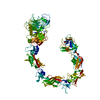
| Entry | Database: PDB / ID: 1av1 | ||||||
|---|---|---|---|---|---|---|---|
| Title | CRYSTAL STRUCTURE OF HUMAN APOLIPOPROTEIN A-I | ||||||
 Components Components | APOLIPOPROTEIN A-I Apolipoprotein AI Apolipoprotein AI | ||||||
 Keywords Keywords | LIPID TRANSPORT / LIPOPROTEIN / CHOLESTEROL METABOLISM / ATHEROSCLEROSIS / HDL / LCAT-ACTIVATION | ||||||
| Function / homology |  Function and homology information Function and homology informationDefective ABCA1 causes TGD / high-density lipoprotein particle receptor binding / Scavenging by Class B Receptors / HDL clearance / spherical high-density lipoprotein particle / positive regulation of hydrolase activity / regulation of intestinal cholesterol absorption / negative regulation of response to cytokine stimulus / protein oxidation / vitamin transport ...Defective ABCA1 causes TGD / high-density lipoprotein particle receptor binding / Scavenging by Class B Receptors / HDL clearance / spherical high-density lipoprotein particle / positive regulation of hydrolase activity / regulation of intestinal cholesterol absorption / negative regulation of response to cytokine stimulus / protein oxidation / vitamin transport / high-density lipoprotein particle binding / cholesterol import / ABC transporters in lipid homeostasis / negative regulation of heterotypic cell-cell adhesion / blood vessel endothelial cell migration / apolipoprotein receptor binding / negative regulation of cell adhesion molecule production / negative regulation of cytokine production involved in immune response / apolipoprotein A-I receptor binding / HDL assembly / peptidyl-methionine modification / negative regulation of very-low-density lipoprotein particle remodeling / phosphatidylcholine biosynthetic process / glucocorticoid metabolic process / Chylomicron remodeling / phosphatidylcholine-sterol O-acyltransferase activator activity / phosphatidylcholine metabolic process / positive regulation of phospholipid efflux / lipid storage / phospholipid homeostasis / Chylomicron assembly / positive regulation of cholesterol metabolic process / high-density lipoprotein particle remodeling / high-density lipoprotein particle clearance / phospholipid efflux / cholesterol transfer activity / reverse cholesterol transport / cholesterol transport / chemorepellent activity / high-density lipoprotein particle assembly / very-low-density lipoprotein particle / positive regulation of CoA-transferase activity / lipoprotein biosynthetic process / high-density lipoprotein particle / endothelial cell proliferation / regulation of Cdc42 protein signal transduction / triglyceride homeostasis / HDL remodeling / cholesterol efflux / Scavenging by Class A Receptors / cholesterol binding / negative regulation of interleukin-1 beta production / positive regulation of Rho protein signal transduction / negative chemotaxis / adrenal gland development / cholesterol biosynthetic process / endocytic vesicle / positive regulation of cholesterol efflux / negative regulation of tumor necrosis factor-mediated signaling pathway / Scavenging of heme from plasma / positive regulation of phagocytosis / positive regulation of substrate adhesion-dependent cell spreading / Retinoid metabolism and transport / positive regulation of stress fiber assembly / endocytic vesicle lumen / heat shock protein binding / cholesterol metabolic process / cholesterol homeostasis / integrin-mediated signaling pathway / Post-translational protein phosphorylation / regulation of protein phosphorylation / phospholipid binding / Heme signaling / PPARA activates gene expression / negative regulation of inflammatory response / Regulation of Insulin-like Growth Factor (IGF) transport and uptake by Insulin-like Growth Factor Binding Proteins (IGFBPs) / extracellular vesicle / Platelet degranulation / amyloid-beta binding / cytoplasmic vesicle / collagen-containing extracellular matrix / secretory granule lumen / blood microparticle / protein stabilization / early endosome / G protein-coupled receptor signaling pathway / Amyloid fiber formation / endoplasmic reticulum lumen / signaling receptor binding / enzyme binding / protein homodimerization activity / extracellular space / extracellular exosome / extracellular region / identical protein binding / plasma membrane / cytosolSimilarity search - Function | ||||||
| Biological species |  Homo sapiens (human) Homo sapiens (human) | ||||||
| Method | X-RAY DIFFRACTION / MIRAS / Resolution: 4 Å | ||||||
 Authors Authors | Borhani, D.W. / Rogers, D.P. / Engler, J.A. / Brouillette, C.G. | ||||||
 Citation Citation | Journal: Proc.Natl.Acad.Sci.USA / Year: 1997 Title: Crystal structure of truncated human apolipoprotein A-I suggests a lipid-bound conformation. Authors: Borhani, D.W. / Rogers, D.P. / Engler, J.A. / Brouillette, C.G. #1: Journal: Biochemistry / Year: 1997Title: Structural Analysis of Apolipoprotein A-I: Limited Proteolysis of Methionine-Reduced and-Oxidized Lipid-Free and Lipid-Bound Human Apo A-I Authors: Roberts, L.M. / Ray, M.J. / Shih, T.W. / Hayden, E. / Reader, M.M. / Brouillette, C.G. #2: Journal: Biochemistry / Year: 1997Title: Truncation of the Amino Terminus of Human Apolipoprotein A-I Substantially Alters Only the Lipid-Free Conformation Authors: Rogers, D.P. / Brouillette, C.G. / Engler, J.A. / Tendian, S.W. / Roberts, L. / Mishra, V.K. / Anantharamaiah, G.M. / Lund-Katz, S. / Phillips, M.C. / Ray, M.J. #3: Journal: Biochim.Biophys.Acta / Year: 1995Title: Structural Models of Human Apolipoprotein A-I Authors: Brouillette, C.G. / Anantharamaiah, G.M. | ||||||
| History |
|
- Structure visualization
| Structure viewer | Molecule: MolmilJmol/JSmol |
|---|
- Downloads & links
-Download
| PDBx/mmCIF format | 1av1.cif.gz | 147 KB | Display | PDBx/mmCIF format |
|---|---|---|---|---|
| PDB format | pdb1av1.ent.gz | 117.6 KB | Display | PDB format |
| PDBx/mmJSON format | 1av1.json.gz | Tree view | PDBx/mmJSON format | |
| Others |  Other downloads Other downloads |
-Validation report
| Arichive directory | https://data.pdbj.org/pub/pdb/validation_reports/av/1av1ftp://data.pdbj.org/pub/pdb/validation_reports/av/1av1 | HTTPS FTP |
|---|
-Related structure data
| Similar structure data |
|---|

-Links
 PDBj
PDBj


- Assembly
| Deposited unit | 
| ||||||||||||||||
|---|---|---|---|---|---|---|---|---|---|---|---|---|---|---|---|---|---|
| 1 |
| ||||||||||||||||
| Unit cell |
| ||||||||||||||||
| Noncrystallographic symmetry (NCS) | NCS oper:
|
-Components
| #1: Protein | Apolipoprotein AI / APO A-I Mass: 23440.559 Da / Num. of mol.: 4 / Fragment: LIPID-BINDING DOMAIN / Mutation: N-TERMINAL MET, DEL(1-43) Source method: isolated from a genetically manipulated source Source: (gene. exp.) Homo sapiens (human) / Tissue: BLOOD / Cell line: BL21 / Plasmid: BL21 / Species (production host): Escherichia coli / Production host:  Escherichia coli BL21(DE3) (bacteria) / Strain (production host): BL21 (DE3) / References: UniProt: P02647 Escherichia coli BL21(DE3) (bacteria) / Strain (production host): BL21 (DE3) / References: UniProt: P02647 |
|---|
-Experimental details
-Experiment
| Experiment | Method: X-RAY DIFFRACTION / Number of used crystals: 2 |
|---|
- Sample preparation
| Crystal | Density Matthews: 5.83 Å3/Da / Density % sol: 72 % Description: DATA WERE EXCLUDED ON A RESOLUTION-DEPENDENT BASIS FOR ALL RESOLUTION SHELLS IN WHICH THE FOR FULLY-RECORDED REFLECTIONS IN THAT SHELL WAS LESS THAN 2.5. Crystal grow | Temperature: 277 K / pH: 7.5 | Details: PROTEIN WAS CRYSTALLIZED FROM 1.2 M NA CITRATE, 100 MM HEPES, PH 7.5 AT 4 DEGREES CELSIUS. CRYSTALS WERE STABILIZED IN 1.4 M NA CITRATE, 100 MM HEPES, PH 7.5., temperature 277K Crystal grow | *PLUS Temperature: 4 ℃ / Method: unknown / PH range low: 7.5 / PH range high: 6.5 Components of the solutions | *PLUS Conc.: 1.2 M / Common name: sodium citrate |
|---|
-Data collection
| Diffraction | Mean temperature: 283 K |
|---|---|
| Diffraction source | Source: ROTATING ANODE / Type: MACSCIENCE / Wavelength: 1.5418 |
| Detector | Type: MARRESEARCH / Detector: IMAGE PLATE / Date: Jan 24, 1996 / Details: DUAL SLITS |
| Radiation | Monochromator: GRAPHITE(002) / Monochromatic (M) / Laue (L): M / Scattering type: x-ray |
| Radiation wavelength | Wavelength: 1.5418 Å / Relative weight: 1 |
| Reflection | Resolution: 4→30 Å / Num. obs: 16089 / % possible obs: 85 % / Observed criterion σ(I): 0 / Redundancy: 3.6 % / Biso Wilson estimate: 70 Å2 / Rmerge(I) obs: 0.166 / Rsym value: 0.166 / Net I/σ(I): 3.7 |
| Reflection shell | Resolution: 4→4.22 Å / Redundancy: 1.3 % / Rmerge(I) obs: 0.732 / Mean I/σ(I) obs: 0.9 / Rsym value: 0.732 / % possible all: 50 |
| Reflection shell | *PLUS % possible obs: 50 % |
- Processing
| Software |
| ||||||||||||||||||||||||||||||||||||||||||||||||||||||||||||
|---|---|---|---|---|---|---|---|---|---|---|---|---|---|---|---|---|---|---|---|---|---|---|---|---|---|---|---|---|---|---|---|---|---|---|---|---|---|---|---|---|---|---|---|---|---|---|---|---|---|---|---|---|---|---|---|---|---|---|---|---|---|
| Refinement | Method to determine structure: MIRAS / Resolution: 4→27 Å / Rfactor Rfree error: 0.015 / Data cutoff high absF: 10000000 / Data cutoff low absF: 0.001 / Isotropic thermal model: 24 (GROUPED) THERMAL FACT / Cross valid method: THROUGHOUT / σ(F): 2 Details: DATA USED IN REFINEMENT WERE SHARPENED BY APPLICATION OF AN ARTIFICIAL TEMPERATURE FACTOR (-70. A**2).
| ||||||||||||||||||||||||||||||||||||||||||||||||||||||||||||
| Displacement parameters |
| ||||||||||||||||||||||||||||||||||||||||||||||||||||||||||||
| Refinement step | Cycle: LAST / Resolution: 4→27 Å
| ||||||||||||||||||||||||||||||||||||||||||||||||||||||||||||
| Refine LS restraints |
| ||||||||||||||||||||||||||||||||||||||||||||||||||||||||||||
| Refine LS restraints NCS | NCS model details: CONSTRAINTS | ||||||||||||||||||||||||||||||||||||||||||||||||||||||||||||
| LS refinement shell | Resolution: 4→4.18 Å / Rfactor Rfree error: 0.064 / Total num. of bins used: 8
| ||||||||||||||||||||||||||||||||||||||||||||||||||||||||||||
| Xplor file |
| ||||||||||||||||||||||||||||||||||||||||||||||||||||||||||||
| Software | *PLUS Name: X-PLOR / Version: 3.843 / Classification: refinement | ||||||||||||||||||||||||||||||||||||||||||||||||||||||||||||
| Refine LS restraints | *PLUS
| ||||||||||||||||||||||||||||||||||||||||||||||||||||||||||||
| LS refinement shell | *PLUS Rfactor Rwork: 0.43 |
