 Movie
Movie Controller

[English] 日本語
 Yorodumi
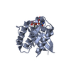
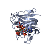
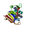
- PDB-1aqz: CRYSTAL STRUCTURE OF A HIGHLY SPECIFIC ASPERGILLUS RIBOTOXIN, RES... -
+ Open data

- Basic information
| Entry | Database: PDB / ID: 1aqz | ||||||
|---|---|---|---|---|---|---|---|
| Title | CRYSTAL STRUCTURE OF A HIGHLY SPECIFIC ASPERGILLUS RIBOTOXIN, RESTRICTOCIN | ||||||
 Components Components | RESTRICTOCIN | ||||||
 Keywords Keywords | RIBOTOXIN /  RIBOSOME-INACTIVATING PROTEIN / PROTEIN-RNA SPECIFIC INTERACTION / LAUE DIFFRACTION / CELL-ENTRY ACTIVITY RIBOSOME-INACTIVATING PROTEIN / PROTEIN-RNA SPECIFIC INTERACTION / LAUE DIFFRACTION / CELL-ENTRY ACTIVITY | ||||||
| Function / homology |  Function and homology informationHydrolases; Acting on ester bonds; Endoribonucleases producing 3'-phosphomonoesters / RNA nuclease activity / negative regulation of translation / RNA binding / extracellular region Function and homology informationHydrolases; Acting on ester bonds; Endoribonucleases producing 3'-phosphomonoesters / RNA nuclease activity / negative regulation of translation / RNA binding / extracellular regionSimilarity search - Function | ||||||
| Biological species |  Aspergillus restrictus (mold) Aspergillus restrictus (mold) | ||||||
| Method | X-RAY DIFFRACTION / SYNCHROTRON / SINGLE ISOMORPHOUS REPLACEMENT, ANOMALOUS SCATTERING / Resolution: 1.7 Å | ||||||
 Authors Authors | Yang, X. / Moffat, K. | ||||||
 Citation Citation | Journal: Structure / Year: 1996 Title: Insights into specificity of cleavage and mechanism of cell entry from the crystal structure of the highly specific Aspergillus ribotoxin, restrictocin. Authors: Yang, X. / Moffat, K. #1: Journal: Proc.Natl.Acad.Sci.USA / Year: 1993Title: The Conformation of the Sarcin/Ricin Loop from 28S Ribosomal RNA Authors: Szewczak, A.A. / Moore, P.B. / Chan, Y.L. / Wool, I.G. #2: Journal: Acta Crystallogr.,Sect.B / Year: 1991Title: Determination and Restrained Least-Squares Refinement of the Structures of Ribonuclease Sa and its Complex with 3'-Guanylic Acid at 1.8 A Resolution Authors: Sevcik, J. / Dodson, E.J. / Dodson, G.G. #3: Journal: J.Mol.Biol. / Year: 1991Title: Crystallization and Preliminary Characterization of Mitogillin, a Ribosomal Ribonuclease from Aspergillus Restrictus Authors: Martinez, S.E. / Smith, J.L. #4: Journal: Trends Biochem.Sci. / Year: 1984Title: The Mechanism of Action of the Cytotoxic Nuclease Alpha-Sarcin and its Use to Analyse Ribosome Structure Authors: Wool, I.G. #5: Journal: Nature / Year: 1982Title: Specific Protein-Nucleic Acid Recognition in Ribonuclease T1-2'-Guanylic Acid Complex: An X-Ray Study Authors: Heinemann, U. / Saenger, W. | ||||||
| History |
|
- Structure visualization
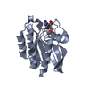
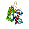
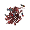
| Structure viewer | Molecule: MolmilJmol/JSmol |
|---|
- Downloads & links
-Download
| PDBx/mmCIF format | 1aqz.cif.gz | 69.5 KB | Display | PDBx/mmCIF format |
|---|---|---|---|---|
| PDB format | pdb1aqz.ent.gz | 56.3 KB | Display | PDB format |
| PDBx/mmJSON format | 1aqz.json.gz | Tree view | PDBx/mmJSON format | |
| Others |  Other downloads Other downloads |
-Validation report
| Arichive directory | https://data.pdbj.org/pub/pdb/validation_reports/aq/1aqzftp://data.pdbj.org/pub/pdb/validation_reports/aq/1aqz | HTTPS FTP |
|---|
-Related structure data
| Similar structure data |
|---|


-Links
 PDBj
PDBj- Assembly
| Deposited unit | 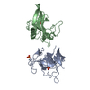
| ||||||||
|---|---|---|---|---|---|---|---|---|---|
| 1 | 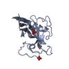
| ||||||||
| 2 | 
| ||||||||
| Unit cell |
| ||||||||
| Noncrystallographic symmetry (NCS) | NCS oper: (Code: given Matrix: (-0.996739, 0.022599, -0.077458), Vector : |
-Components
| #1: Protein | Mass: 16888.893 Da / Num. of mol.: 2 / Source method: isolated from a natural source / Details: INORGANIC PHOSPHATE GROUP AT THE ACTIVE SITE / Source: (natural) Aspergillus restrictus (mold)References: UniProt: P67876, Hydrolases; Acting on ester bonds; Endoribonucleases producing 3'-phosphomonoesters#2: Chemical | Phosphate  Mass: 94.971 Da / Num. of mol.: 3 / Source method: obtained synthetically / Formula: PO4 Mass: 94.971 Da / Num. of mol.: 3 / Source method: obtained synthetically / Formula: PO4#3: Water | ChemComp-HOH / | Water Mass: 18.015 Da / Num. of mol.: 202 / Source method: isolated from a natural source / Formula: H2O Mass: 18.015 Da / Num. of mol.: 202 / Source method: isolated from a natural source / Formula: H2O |
|---|
-Experimental details
-Experiment
| Experiment | Method: X-RAY DIFFRACTION / Number of used crystals: 1 |
|---|
- Sample preparation
| Crystal | Density Matthews: 2.29 Å3/Da / Density % sol: 46 % Description: DATA WERE COLLECTED USING LAUE DIFFRACTION. 62 IMAGES WERE COLLECTED. TOTAL EXPOSURE TIME IS 325 MS. | ||||||||||||||||||||
|---|---|---|---|---|---|---|---|---|---|---|---|---|---|---|---|---|---|---|---|---|---|
| Crystal grow | Method: vapor diffusion, microdialysis / pH: 6.8 Details: A COMBINATION OF VAPOR DIFFUSION AND MICRODIALYSIS TECHNIQUES WERE USED TO CRYSTALLIZE RESTRICTOCIN. THE FINAL MOTHER LIQUOR CONSISTS OF 60% ETHANOL, 10MM SODIUM PHOSPHATE, PH6.8. THE ...Details: A COMBINATION OF VAPOR DIFFUSION AND MICRODIALYSIS TECHNIQUES WERE USED TO CRYSTALLIZE RESTRICTOCIN. THE FINAL MOTHER LIQUOR CONSISTS OF 60% ETHANOL, 10MM SODIUM PHOSPHATE, PH6.8. THE PROTEIN CONCENTRATION IS 10MG/ML., vapor diffusion and microdialysis | ||||||||||||||||||||
| Crystal grow | *PLUS Method: vapor diffusion | ||||||||||||||||||||
| Components of the solutions | *PLUS
|
-Data collection
| Diffraction | Mean temperature: 298 K | |||||||||
|---|---|---|---|---|---|---|---|---|---|---|
| Diffraction source | Source: SYNCHROTRON / Site: NSLS  / Beamline: X26C / Wavelength: 0.7 / Wavelength: 0.7, 2.05 / Beamline: X26C / Wavelength: 0.7 / Wavelength: 0.7, 2.05 | |||||||||
| Detector | Type: FUJI / Detector: IMAGE PLATE / Date: Aug 1, 1994 / Details: MIRROR | |||||||||
| Radiation | Monochromatic (M) / Laue (L): L / Scattering type: x-ray | |||||||||
| Radiation wavelength |
| |||||||||
| Reflection | Resolution: 1.7→8 Å / Num. obs: 28328 / % possible obs: 85.6 % / Observed criterion σ(I): 2 / Redundancy: 8 % / Rmerge(I) obs: 0.039 | |||||||||
| Reflection shell | Resolution: 1.7→1.78 Å / % possible all: 54.8 |
- Processing
| Software |
| ||||||||||||||||||||||||||||||||||||||||||||||||||||||||||||
|---|---|---|---|---|---|---|---|---|---|---|---|---|---|---|---|---|---|---|---|---|---|---|---|---|---|---|---|---|---|---|---|---|---|---|---|---|---|---|---|---|---|---|---|---|---|---|---|---|---|---|---|---|---|---|---|---|---|---|---|---|---|
| Refinement | Method to determine structure: SINGLE ISOMORPHOUS REPLACEMENT, ANOMALOUS SCATTERING Resolution: 1.7→8 Å / Cross valid method: THROUGHOUT / σ(F): 2
| ||||||||||||||||||||||||||||||||||||||||||||||||||||||||||||
| Displacement parameters | Biso mean: 19.9 Å2 | ||||||||||||||||||||||||||||||||||||||||||||||||||||||||||||
| Refine analyze |
| ||||||||||||||||||||||||||||||||||||||||||||||||||||||||||||
| Refinement step | Cycle: LAST / Resolution: 1.7→8 Å
| ||||||||||||||||||||||||||||||||||||||||||||||||||||||||||||
| Refine LS restraints |
| ||||||||||||||||||||||||||||||||||||||||||||||||||||||||||||
| LS refinement shell | Resolution: 1.7→1.78 Å / Total num. of bins used: 8
| ||||||||||||||||||||||||||||||||||||||||||||||||||||||||||||
| Xplor file |
| ||||||||||||||||||||||||||||||||||||||||||||||||||||||||||||
| Software | *PLUS Name: X-PLOR / Version: 3.1 / Classification: refinement | ||||||||||||||||||||||||||||||||||||||||||||||||||||||||||||
| Refinement | *PLUS Rfactor obs: 0.177 / Rfactor Rfree: 0.237 | ||||||||||||||||||||||||||||||||||||||||||||||||||||||||||||
| Solvent computation | *PLUS | ||||||||||||||||||||||||||||||||||||||||||||||||||||||||||||
| Displacement parameters | *PLUS | ||||||||||||||||||||||||||||||||||||||||||||||||||||||||||||
| Refine LS restraints | *PLUS
|
