 Movie
Movie Controller

[English] 日本語
 Yorodumi
- PDB-1ad3: CLASS 3 ALDEHYDE DEHYDROGENASE COMPLEX WITH NICOTINAMIDE-ADENINE-... -
+ Open data

- Basic information
| Entry | Database: PDB / ID: 1ad3 | ||||||
|---|---|---|---|---|---|---|---|
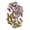
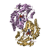
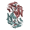
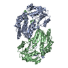
| Title | CLASS 3 ALDEHYDE DEHYDROGENASE COMPLEX WITH NICOTINAMIDE-ADENINE-DINUCLEOTIDE | ||||||
 Components Components | ALDEHYDE DEHYDROGENASE (CLASS 3) | ||||||
 Keywords Keywords |  OXIDOREDUCTASE / NADP / AROMATIC ALDEHYDE OXIDOREDUCTASE / NADP / AROMATIC ALDEHYDE | ||||||
| Function / homology |  Function and homology information Function and homology informationaldehyde dehydrogenase [NAD(P)+] / Phase I - Functionalization of compounds / 3-chloroallyl aldehyde dehydrogenase activity / benzaldehyde dehydrogenase (NAD+) activity / alcohol dehydrogenase (NADP+) activity / : / cellular aldehyde metabolic process / response to xenobiotic stimulus => GO:0009410 / : / oxidoreductase activity, acting on the aldehyde or oxo group of donors, NAD or NADP as acceptor ...aldehyde dehydrogenase [NAD(P)+] / Phase I - Functionalization of compounds / 3-chloroallyl aldehyde dehydrogenase activity / benzaldehyde dehydrogenase (NAD+) activity / alcohol dehydrogenase (NADP+) activity / : / cellular aldehyde metabolic process / response to xenobiotic stimulus => GO:0009410 / : / oxidoreductase activity, acting on the aldehyde or oxo group of donors, NAD or NADP as acceptor / aldehyde dehydrogenase (NAD+) activity / response to glucocorticoid / response to cAMP / response to nutrient / response to organic cyclic compound / membrane => GO:0016020 / response to hypoxia / positive regulation of cell population proliferation / endoplasmic reticulum / plasma membrane / cytosol / cytoplasmSimilarity search - Function | ||||||
| Biological species |  Rattus norvegicus (Norway rat) Rattus norvegicus (Norway rat) | ||||||
| Method | X-RAY DIFFRACTION / ISIRAS / Resolution: 2.6 Å | ||||||
 Authors Authors | Liu, Z.-J. / Rose, J. / Wang, B.C. | ||||||
 Citation Citation | Journal: Nat.Struct.Biol. / Year: 1997 Title: The first structure of an aldehyde dehydrogenase reveals novel interactions between NAD and the Rossmann fold. Authors: Liu, Z.J. / Sun, Y.J. / Rose, J. / Chung, Y.J. / Hsiao, C.D. / Chang, W.R. / Kuo, I. / Perozich, J. / Lindahl, R. / Hempel, J. / Wang, B.C. #1: Journal: Enzymology and Molecular Biology of Carbonyl Metabolism 6 (in: Adv.Exp. Med.Biol., V.414)Year: 1997 Title: Crystal Structure of a Class 3 Aldehyde Dehydrogenase at 2.6 Angstroms Resolution Authors: Liu, Z.-J. / Hempel, J. / Sun, J. / Rose, J. / Hsiao, D. / Chang, W.-R. / Chung, Y.-J. / Kuo, I. / Lindahl, R. / Wang, B.-C. #2: Journal: Enzymology and Molecular Biology of Carbonyl Metabolism 6 (in: Adv.Exp. Med.Biol., V.414)Year: 1997 Title: Conserved Residues in the Aldehyde Dehydrogenase Family Authors: Hempel, J. / Liu, Z.-J. / Perozich, J. / Rose, J. / Lindahl, R. / Wang, B.-C. #3: Journal: Proteins / Year: 1990Title: Preliminary Crystallographic Analysis of Class 3 Rat Liver Aldehyde Dehydrogenase Authors: Rose, J.P. / Hempel, J. / Kuo, I. / Lindahl, R. / Wang, B.C. | ||||||
| History |
|
- Structure visualization
| Structure viewer | Molecule: MolmilJmol/JSmol |
|---|
- Downloads & links
-Download
| PDBx/mmCIF format | 1ad3.cif.gz | 216.7 KB | Display | PDBx/mmCIF format |
|---|---|---|---|---|
| PDB format | pdb1ad3.ent.gz | 176.4 KB | Display | PDB format |
| PDBx/mmJSON format | 1ad3.json.gz | Tree view | PDBx/mmJSON format | |
| Others |  Other downloads Other downloads |
-Validation report
| Arichive directory | https://data.pdbj.org/pub/pdb/validation_reports/ad/1ad3ftp://data.pdbj.org/pub/pdb/validation_reports/ad/1ad3 | HTTPS FTP |
|---|
-Related structure data
| Similar structure data |
|---|


-Links
 PDBj
PDBj

- Assembly
| Deposited unit | 
| ||||||||
|---|---|---|---|---|---|---|---|---|---|
| 1 |
| ||||||||
| Unit cell |
| ||||||||
| Noncrystallographic symmetry (NCS) | NCS oper: (Code: given Matrix: (-0.1895, 0.0654, -0.9797), Vector : Details | ALDEHYDE DEHYDROGENASE IS A HOMODIMER. THE FULL DIMER IS PRESENT IN THE CRYSTALLOGRAPHIC ASYMMETRIC UNIT. | |
-Components
| #1: Protein | Mass: 50313.438 Da / Num. of mol.: 2 Source method: isolated from a genetically manipulated source Source: (gene. exp.) Rattus norvegicus (Norway rat) / Organ: LIVER / Plasmid: PTA1DH / Production host:  Escherichia coli (E. coli) / Strain (production host): BH101 Escherichia coli (E. coli) / Strain (production host): BH101References: UniProt: P11883, aldehyde dehydrogenase [NAD(P)+] #2: Chemical | Nicotinamide adenine dinucleotide  Mass: 663.425 Da / Num. of mol.: 2 / Source method: obtained synthetically / Formula: C21H27N7O14P2 / Comment: NAD*YM Mass: 663.425 Da / Num. of mol.: 2 / Source method: obtained synthetically / Formula: C21H27N7O14P2 / Comment: NAD*YM#3: Water | ChemComp-HOH / | Water Mass: 18.015 Da / Num. of mol.: 276 / Source method: isolated from a natural source / Formula: H2O Mass: 18.015 Da / Num. of mol.: 276 / Source method: isolated from a natural source / Formula: H2O |
|---|
-Experimental details
-Experiment
| Experiment | Method: X-RAY DIFFRACTION / Number of used crystals: 1 |
|---|
- Sample preparation
| Crystal | Density Matthews: 2.45 Å3/Da / Density % sol: 50 % | ||||||||||||||||||||||||||||||||||||||||||||||||||||||||||||
|---|---|---|---|---|---|---|---|---|---|---|---|---|---|---|---|---|---|---|---|---|---|---|---|---|---|---|---|---|---|---|---|---|---|---|---|---|---|---|---|---|---|---|---|---|---|---|---|---|---|---|---|---|---|---|---|---|---|---|---|---|---|
| Crystal grow | pH: 6.2 / Details: pH 6.2 | ||||||||||||||||||||||||||||||||||||||||||||||||||||||||||||
| Crystal grow | *PLUS Method: vapor diffusionDetails: Rose, J.P., (1990) Proteins: Struct.,Funct., Genet., 8, 305. | ||||||||||||||||||||||||||||||||||||||||||||||||||||||||||||
| Components of the solutions | *PLUS
|
-Data collection
| Diffraction | Mean temperature: 289 K |
|---|---|
| Diffraction source | Source: ROTATING ANODE / Type: RIGAKU RUH2R / Wavelength: 1.5418 |
| Detector | Type: SIEMENS / Detector: AREA DETECTOR / Date: Jun 1, 1994 / Details: SUPPER MIRRORS (SMALL) |
| Radiation | Monochromator: NI FILTER / Monochromatic (M) / Laue (L): M / Scattering type: x-ray |
| Radiation wavelength | Wavelength: 1.5418 Å / Relative weight: 1 |
| Reflection | Highest resolution: 2.5 Å / Num. obs: 25656 / % possible obs: 80 % / Observed criterion σ(I): -3 / Redundancy: 2.3 % / Biso Wilson estimate: 23.62 Å2 / Rmerge(I) obs: 0.05 / Net I/σ(I): 17.444 |
| Reflection shell | Resolution: 2.6→2.7 Å / Redundancy: 2.3 % / Rmerge(I) obs: 0.183 / Mean I/σ(I) obs: 2.29 / % possible all: 50 |
| Reflection shell | *PLUS % possible obs: 50 % |
- Processing
| Software |
| ||||||||||||||||||||||||||||||||||||||||||||||||||||||||||||
|---|---|---|---|---|---|---|---|---|---|---|---|---|---|---|---|---|---|---|---|---|---|---|---|---|---|---|---|---|---|---|---|---|---|---|---|---|---|---|---|---|---|---|---|---|---|---|---|---|---|---|---|---|---|---|---|---|---|---|---|---|---|
| Refinement | Method to determine structure: ISIRAS / Resolution: 2.6→8 Å / Rfactor Rfree error: 0.006 / Cross valid method: THROUGHOUT / σ(F): 2
| ||||||||||||||||||||||||||||||||||||||||||||||||||||||||||||
| Displacement parameters | Biso mean: 18.4 Å2 | ||||||||||||||||||||||||||||||||||||||||||||||||||||||||||||
| Refine analyze | Luzzati coordinate error obs: 0.35 Å | ||||||||||||||||||||||||||||||||||||||||||||||||||||||||||||
| Refinement step | Cycle: LAST / Resolution: 2.6→8 Å
| ||||||||||||||||||||||||||||||||||||||||||||||||||||||||||||
| Refine LS restraints |
| ||||||||||||||||||||||||||||||||||||||||||||||||||||||||||||
| LS refinement shell | Resolution: 2.6→2.7 Å / Rfactor Rfree error: 0.031
| ||||||||||||||||||||||||||||||||||||||||||||||||||||||||||||
| Xplor file |
| ||||||||||||||||||||||||||||||||||||||||||||||||||||||||||||
| Software | *PLUS Name: X-PLOR / Version: 3.1 / Classification: refinement | ||||||||||||||||||||||||||||||||||||||||||||||||||||||||||||
| Refinement | *PLUS Rfactor all: 0.192 | ||||||||||||||||||||||||||||||||||||||||||||||||||||||||||||
| Solvent computation | *PLUS | ||||||||||||||||||||||||||||||||||||||||||||||||||||||||||||
| Displacement parameters | *PLUS | ||||||||||||||||||||||||||||||||||||||||||||||||||||||||||||
| Refine LS restraints | *PLUS
|
