 Movie
Movie Controller

[English] 日本語
 Yorodumi
- PDB-1abo: CRYSTAL STRUCTURE OF THE COMPLEX OF THE ABL TYROSINE KINASE SH3 D... -
+ Open data

- Basic information
| Entry | Database: PDB / ID: 1abo | ||||||
|---|---|---|---|---|---|---|---|
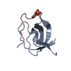
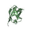
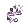
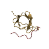
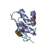
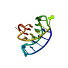
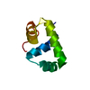
| Title | CRYSTAL STRUCTURE OF THE COMPLEX OF THE ABL TYROSINE KINASE SH3 DOMAIN WITH 3BP-1 SYNTHETIC PEPTIDE | ||||||
 Components Components |
| ||||||
 Keywords Keywords | COMPLEX (KINASE/PEPTIDE) /  SH3 DOMAIN / TRANSFERASE (PHOSPHOTRANSFERASE) / PROTO-ONCOGENE / COMPLEX (KINASE-PEPTIDE) COMPLEX SH3 DOMAIN / TRANSFERASE (PHOSPHOTRANSFERASE) / PROTO-ONCOGENE / COMPLEX (KINASE-PEPTIDE) COMPLEX | ||||||
| Function / homology |  Function and homology information Function and homology informationregulation of actin filament depolymerization / small GTPase binding => GO:0031267 / Role of ABL in ROBO-SLIT signaling / HDR through Single Strand Annealing (SSA) / negative regulation of small GTPase mediated signal transduction / RHO GTPases Activate WASPs and WAVEs / semaphorin receptor binding / Cyclin D associated events in G1 / Recruitment and ATM-mediated phosphorylation of repair and signaling proteins at DNA double strand breaks / transitional one stage B cell differentiation ...regulation of actin filament depolymerization / small GTPase binding => GO:0031267 / Role of ABL in ROBO-SLIT signaling / HDR through Single Strand Annealing (SSA) / negative regulation of small GTPase mediated signal transduction / RHO GTPases Activate WASPs and WAVEs / semaphorin receptor binding / Cyclin D associated events in G1 / Recruitment and ATM-mediated phosphorylation of repair and signaling proteins at DNA double strand breaks / transitional one stage B cell differentiation / protein localization to cytoplasmic microtubule plus-end / DNA conformation change / circulatory system development / podocyte apoptotic process / DN4 thymocyte differentiation / RUNX1 regulates transcription of genes involved in differentiation of HSCs / response to epinephrine / regulation of cellular senescence / ruffle assembly / regulation of modification of synaptic structure / regulation of Rac protein signal transduction / delta-catenin binding / B cell proliferation involved in immune response / positive regulation of extracellular matrix organization / Regulation of actin dynamics for phagocytic cup formation / neuroepithelial cell differentiation / microspike assembly / positive regulation of Wnt signaling pathway, planar cell polarity pathway / regulation of extracellular matrix organization / cerebellum morphogenesis / positive regulation of blood vessel branching / regulation of blood vessel endothelial cell migration / B-1 B cell homeostasis / neuropilin signaling pathway / neuropilin binding / Myogenesis / bubble DNA binding / cell junction assembly / filopodium assembly / activated T cell proliferation / establishment of epithelial cell apical/basal polarity / regulation of Cdc42 protein signal transduction / proline-rich region binding / positive regulation of dendrite development / mitogen-activated protein kinase binding / myoblast proliferation / alpha-beta T cell differentiation / syntaxin binding / cardiac muscle cell proliferation / regulation of T cell differentiation / regulation of axon extension / phagocytic cup / phagocytosis, engulfment / negative regulation of double-strand break repair via homologous recombination / positive regulation of cell migration involved in sprouting angiogenesis / negative regulation of cell-cell adhesion / regulation of microtubule polymerization / B cell proliferation / positive regulation of osteoblast proliferation / cell leading edge / platelet-derived growth factor receptor-beta signaling pathway / positive regulation of focal adhesion assembly / negative regulation of cellular senescence / Bergmann glial cell differentiation / associative learning / semaphorin-plexin signaling pathway / neuromuscular process controlling balance / platelet-derived growth factor receptor signaling pathway / negative regulation of BMP signaling pathway / negative regulation of mitotic cell cycle / negative regulation of long-term synaptic potentiation / endothelial cell migration / bicellular tight junction / positive regulation of T cell migration / canonical NF-kappaB signal transduction / BMP signaling pathway / phagocytosis / negative regulation of endothelial cell apoptotic process / four-way junction DNA binding / signal transduction in response to DNA damage / positive regulation of substrate adhesion-dependent cell spreading / positive regulation of vasoconstriction / positive regulation of stress fiber assembly / spleen development / ERK1 and ERK2 cascade / ruffle / cellular response to transforming growth factor beta stimulus / positive regulation of establishment of T cell polarity / actin filament polymerization / positive regulation of interleukin-2 production / phosphotyrosine residue binding / response to endoplasmic reticulum stress / SH2 domain binding / GTPase activator activity / ephrin receptor binding / positive regulation of mitotic cell cycle / substrate adhesion-dependent cell spreading / post-embryonic development / positive regulation of release of sequestered calcium ion into cytosol / positive regulation of endothelial cell migrationSimilarity search - Function | ||||||
| Biological species |  Mus musculus (house mouse) Mus musculus (house mouse) | ||||||
| Method | X-RAY DIFFRACTION / Resolution: 2 Å | ||||||
 Authors Authors | Musacchio, A. / Wilmanns, M. / Saraste, M. | ||||||
 Citation Citation | Journal: Nat.Struct.Biol. / Year: 1994 Title: High-resolution crystal structures of tyrosine kinase SH3 domains complexed with proline-rich peptides. Authors: Musacchio, A. / Saraste, M. / Wilmanns, M. #1: Journal: Embo J. / Year: 1993Title: Crystal Structure of the SH3 Domain in Human Fyn. Comparison of the Three-Dimensional Structures of the SH3 Domain in Tyrosine Kinases and Spectrin Authors: Noble, M.E.M. / Musacchio, A. / Courtneidge, S. / Saraste, M. / Wierenga, R. #2: Journal: Science / Year: 1993Title: Identification of a Ten-Amino Acid SH3 Binding Site Authors: Ren, R. / Mayer, B. / Clark, K.L. / Baltimore, D. #3: Journal: Nature / Year: 1992Title: Crystal Structure of a Src-Homology 3 (SH3) Domain Authors: Musacchio, A. / Noble, M.E.M. / Pauptit, R. / Wierenga, R. / Saraste, M. | ||||||
| History |
|
- Structure visualization
| Structure viewer | Molecule: MolmilJmol/JSmol |
|---|
- Downloads & links
-Download
| PDBx/mmCIF format | 1abo.cif.gz | 40.9 KB | Display | PDBx/mmCIF format |
|---|---|---|---|---|
| PDB format | pdb1abo.ent.gz | 28.7 KB | Display | PDB format |
| PDBx/mmJSON format | 1abo.json.gz | Tree view | PDBx/mmJSON format | |
| Others |  Other downloads Other downloads |
-Validation report
| Arichive directory | https://data.pdbj.org/pub/pdb/validation_reports/ab/1aboftp://data.pdbj.org/pub/pdb/validation_reports/ab/1abo | HTTPS FTP |
|---|
-Related structure data


-Links
 PDBj
PDBj


- Assembly
| Deposited unit | 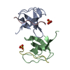
| ||||||||
|---|---|---|---|---|---|---|---|---|---|
| 1 | 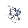
| ||||||||
| 2 | 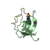
| ||||||||
| 3 | 
| ||||||||
| Unit cell |
|
-Components
| #1: Protein | Mass: 6880.580 Da / Num. of mol.: 2 Source method: isolated from a genetically manipulated source Source: (gene. exp.) Mus musculus (house mouse) / Plasmid: PET / Production host:  Escherichia coli (E. coli) / References: UniProt: P00520, EC: 2.7.1.112 Escherichia coli (E. coli) / References: UniProt: P00520, EC: 2.7.1.112#2: Protein/peptide | Mass: 1017.239 Da / Num. of mol.: 2 Source method: isolated from a genetically manipulated source References: UniProt: P55194 #3: Chemical | Sulfate  Mass: 96.063 Da / Num. of mol.: 2 / Source method: obtained synthetically / Formula: SO4 Mass: 96.063 Da / Num. of mol.: 2 / Source method: obtained synthetically / Formula: SO4#4: Water | ChemComp-HOH / | Water Mass: 18.015 Da / Num. of mol.: 121 / Source method: isolated from a natural source / Formula: H2O Mass: 18.015 Da / Num. of mol.: 121 / Source method: isolated from a natural source / Formula: H2O |
|---|
-Experimental details
-Experiment
| Experiment | Method: X-RAY DIFFRACTION |
|---|
- Sample preparation
| Crystal | Density Matthews: 2.01 Å3/Da / Density % sol: 38.9 % | ||||||||||||||||||||||||||||||||||||||||||||||||||||||||||||||||||
|---|---|---|---|---|---|---|---|---|---|---|---|---|---|---|---|---|---|---|---|---|---|---|---|---|---|---|---|---|---|---|---|---|---|---|---|---|---|---|---|---|---|---|---|---|---|---|---|---|---|---|---|---|---|---|---|---|---|---|---|---|---|---|---|---|---|---|---|
| Crystal grow | *PLUS pH: 8.2 / Method: vapor diffusion, hanging drop / Details: used as seeds | ||||||||||||||||||||||||||||||||||||||||||||||||||||||||||||||||||
| Components of the solutions | *PLUS
|
-Data collection
| Diffraction source | Wavelength: 1.5418 Å |
|---|---|
| Detector | Type: MARRESEARCH / Detector: IMAGE PLATE / Date: Dec 20, 1993 |
| Radiation | Monochromatic (M) / Laue (L): M / Scattering type: x-ray |
| Radiation wavelength | Wavelength: 1.5418 Å / Relative weight: 1 |
| Reflection | % possible obs: 89 % / Rmerge(I) obs: 0.029 |
| Reflection | *PLUS Highest resolution: 2 Å / Rmerge(I) obs: 0.029 |
- Processing
| Software |
| ||||||||||||||||||||||||||||||||||||||||||||||||||||||||||||
|---|---|---|---|---|---|---|---|---|---|---|---|---|---|---|---|---|---|---|---|---|---|---|---|---|---|---|---|---|---|---|---|---|---|---|---|---|---|---|---|---|---|---|---|---|---|---|---|---|---|---|---|---|---|---|---|---|---|---|---|---|---|
| Refinement | Resolution: 2→8 Å / σ(F): 1 /
| ||||||||||||||||||||||||||||||||||||||||||||||||||||||||||||
| Refinement step | Cycle: LAST / Resolution: 2→8 Å
| ||||||||||||||||||||||||||||||||||||||||||||||||||||||||||||
| Refine LS restraints |
|
