 Movie
Movie Controller


+ Open data

- Basic information
| Entry | Database: PDB / ID: 1a9c | ||||||
|---|---|---|---|---|---|---|---|
| Title | GTP CYCLOHYDROLASE I (C110S MUTANT) IN COMPLEX WITH GTP | ||||||
 Components Components | GTP CYCLOHYDROLASE I | ||||||
 Keywords Keywords | HYDROLASE / GTP / PURINE HYDROLYSIS / PTERINE SYNTHESIS / TETRAHYDROBIOPTERIN | ||||||
| Function / homology |  Function and homology informationGTP cyclohydrolase I / GTP cyclohydrolase I activity / : / tetrahydrobiopterin biosynthetic process / queuosine biosynthetic process / tetrahydrofolate biosynthetic process / one-carbon metabolic process / GTP binding / protein-containing complex / zinc ion binding ...GTP cyclohydrolase I / GTP cyclohydrolase I activity / : / tetrahydrobiopterin biosynthetic process / queuosine biosynthetic process / tetrahydrofolate biosynthetic process / one-carbon metabolic process / GTP binding / protein-containing complex / zinc ion binding / identical protein binding / cytosol / cytoplasm Function and homology informationGTP cyclohydrolase I / GTP cyclohydrolase I activity / : / tetrahydrobiopterin biosynthetic process / queuosine biosynthetic process / tetrahydrofolate biosynthetic process / one-carbon metabolic process / GTP binding / protein-containing complex / zinc ion binding ...GTP cyclohydrolase I / GTP cyclohydrolase I activity / : / tetrahydrobiopterin biosynthetic process / queuosine biosynthetic process / tetrahydrofolate biosynthetic process / one-carbon metabolic process / GTP binding / protein-containing complex / zinc ion binding / identical protein binding / cytosol / cytoplasmSimilarity search - Function | ||||||
| Biological species |  Escherichia coli (E. coli) Escherichia coli (E. coli) | ||||||
| Method | X-RAY DIFFRACTION / Resolution: 2.9 Å | ||||||
 Authors Authors | Auerbach, G. / Nar, H. / Bracher, A. / Bacher, A. / Huber, R. | ||||||
 Citation Citation | Journal: To be Published Title: GTP Cyclohydrolase I in Complex with GTP at 2.1 A Resolution Authors: Auerbach, G. / Bracher, A. / Nar, H. / Fischer, M. / Hoesl, C. / Huber, R. / Bacher, A. #1: Journal: Biol.Chem. / Year: 1997Title: The Pathway from GTP to Tetrahydrobiopterin: Three-Dimensional Structures of GTP Cyclohydrolase I and 6-Pyruvoyl Tetrahydropterin Synthase Authors: Auerbach, G. / Nar, H. #2: Journal: Embo J. / Year: 1997Title: The 1.25 A Crystal Structure of Sepiapterin Reductase Reveals its Binding Mode to Pterins and Brain Neurotransmitters Authors: Auerbach, G. / Herrmann, A. / Gutlich, M. / Fischer, M. / Jacob, U. / Bacher, A. / Huber, R. #3: Journal: Proc.Natl.Acad.Sci.USA / Year: 1995Title: Active Site Topology and Reaction Mechanism of GTP Cyclohydrolase I Authors: Nar, H. / Huber, R. / Auerbach, G. / Fischer, M. / Hosl, C. / Ritz, H. / Bracher, A. / Meining, W. / Eberhardt, S. / Bacher, A. #4: Journal: Structure / Year: 1995Title: Atomic Structure of GTP Cyclohydrolase I Authors: Nar, H. / Huber, R. / Meining, W. / Schmid, C. / Weinkauf, S. / Bacher, A. | ||||||
| History |
|
- Structure visualization
| Structure viewer | Molecule: MolmilJmol/JSmol |
|---|
- Downloads & links
-Download
| PDBx/mmCIF format | 1a9c.cif.gz | 784.3 KB | Display | PDBx/mmCIF format |
|---|---|---|---|---|
| PDB format | pdb1a9c.ent.gz | 653.2 KB | Display | PDB format |
| PDBx/mmJSON format | 1a9c.json.gz | Tree view | PDBx/mmJSON format | |
| Others |  Other downloads Other downloads |
-Validation report
| Arichive directory | https://data.pdbj.org/pub/pdb/validation_reports/a9/1a9cftp://data.pdbj.org/pub/pdb/validation_reports/a9/1a9c | HTTPS FTP |
|---|
-Related structure data
| Similar structure data |
|---|


-Links
 PDBj
PDBj
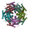
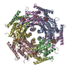
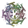
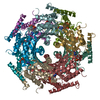
- Assembly
| Deposited unit | 
| ||||||||||||||||||||||||||||||||||||||||||||||||||||||||||||
|---|---|---|---|---|---|---|---|---|---|---|---|---|---|---|---|---|---|---|---|---|---|---|---|---|---|---|---|---|---|---|---|---|---|---|---|---|---|---|---|---|---|---|---|---|---|---|---|---|---|---|---|---|---|---|---|---|---|---|---|---|---|
| 1 | 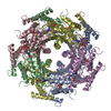
| ||||||||||||||||||||||||||||||||||||||||||||||||||||||||||||
| 2 | 
| ||||||||||||||||||||||||||||||||||||||||||||||||||||||||||||
| Unit cell |
| ||||||||||||||||||||||||||||||||||||||||||||||||||||||||||||
| Noncrystallographic symmetry (NCS) | NCS oper:
|
-Components
| #1: Protein | Mass: 24717.387 Da / Num. of mol.: 15 / Mutation: C110S Source method: isolated from a genetically manipulated source Source: (gene. exp.) Escherichia coli (E. coli) / Description: HOMOLOGOUS EXPRESSION / Organ: BRAIN / Production host: Escherichia coli (E. coli) / References: UniProt: P0A6T5, GTP cyclohydrolase I#2: Chemical | ChemComp-GTP / Guanosine triphosphate  Mass: 523.180 Da / Num. of mol.: 15 / Source method: obtained synthetically / Formula: C10H16N5O14P3 / Comment: GTP, energy-carrying molecule*YM Mass: 523.180 Da / Num. of mol.: 15 / Source method: obtained synthetically / Formula: C10H16N5O14P3 / Comment: GTP, energy-carrying molecule*YM#3: Water | ChemComp-HOH / | Water Mass: 18.015 Da / Num. of mol.: 210 / Source method: isolated from a natural source / Formula: H2O Mass: 18.015 Da / Num. of mol.: 210 / Source method: isolated from a natural source / Formula: H2O |
|---|
-Experimental details
-Experiment
| Experiment | Method: X-RAY DIFFRACTION / Number of used crystals: 1 |
|---|
- Sample preparation
| Crystal | Density Matthews: 3.16 Å3/Da / Density % sol: 61 % |
|---|---|
| Crystal grow | pH: 7 / Details: pH 7.0 |
-Data collection
| Diffraction | Mean temperature: 280 K |
|---|---|
| Diffraction source | Source: ROTATING ANODE / Type: RIGAKU / Wavelength: 1.5418 |
| Detector | Type: MARRESEARCH / Detector: IMAGE PLATE / Date: Jan 6, 1996 |
| Radiation | Monochromatic (M) / Laue (L): M / Scattering type: x-ray |
| Radiation wavelength | Wavelength: 1.5418 Å / Relative weight: 1 |
| Reflection | Resolution: 2.9→25 Å / Num. obs: 86307 / % possible obs: 84.4 % / Observed criterion σ(I): 0 / Rmerge(I) obs: 0.082 |
- Processing
| Software |
| ||||||||||||||||||||||||||||||||||||||||||||||||||||||||||||
|---|---|---|---|---|---|---|---|---|---|---|---|---|---|---|---|---|---|---|---|---|---|---|---|---|---|---|---|---|---|---|---|---|---|---|---|---|---|---|---|---|---|---|---|---|---|---|---|---|---|---|---|---|---|---|---|---|---|---|---|---|---|
| Refinement | Resolution: 2.9→8 Å / Cross valid method: THROUGHOUT / σ(F): 0
| ||||||||||||||||||||||||||||||||||||||||||||||||||||||||||||
| Displacement parameters | Biso mean: 32.6 Å2 | ||||||||||||||||||||||||||||||||||||||||||||||||||||||||||||
| Refinement step | Cycle: LAST / Resolution: 2.9→8 Å
| ||||||||||||||||||||||||||||||||||||||||||||||||||||||||||||
| Refine LS restraints |
|
