 Movie
Movie Controller


+ Open data

- Basic information
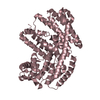
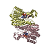
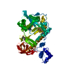
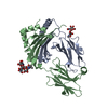
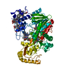
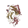
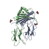
| Entry | Database: PDB / ID: 1a98 | ||||||
|---|---|---|---|---|---|---|---|
| Title | XPRTASE FROM E. COLI COMPLEXED WITH GMP | ||||||
 Components Components | XANTHINE-GUANINE PHOSPHORIBOSYLTRANSFERASE | ||||||
 Keywords Keywords |  PHOSPHORIBOSYLTRANSFERASE / TRANSFERASE / PURINE SALVAGE ENZYME / GLYCOSYLTRANSFERASE PHOSPHORIBOSYLTRANSFERASE / TRANSFERASE / PURINE SALVAGE ENZYME / GLYCOSYLTRANSFERASE | ||||||
| Function / homology |  Function and homology informationxanthine phosphoribosyltransferase / XMP salvage / xanthine phosphoribosyltransferase activity / GMP salvage / guanine phosphoribosyltransferase activity / hypoxanthine phosphoribosyltransferase activity / IMP salvage / purine ribonucleoside salvage / guanosine tetraphosphate binding / Transferases; Glycosyltransferases; Pentosyltransferases ...xanthine phosphoribosyltransferase / XMP salvage / xanthine phosphoribosyltransferase activity / GMP salvage / guanine phosphoribosyltransferase activity / hypoxanthine phosphoribosyltransferase activity / IMP salvage / purine ribonucleoside salvage / guanosine tetraphosphate binding / Transferases; Glycosyltransferases; Pentosyltransferases / protein homotetramerization / magnesium ion binding / protein-containing complex / identical protein binding / plasma membrane / cytosol Function and homology informationxanthine phosphoribosyltransferase / XMP salvage / xanthine phosphoribosyltransferase activity / GMP salvage / guanine phosphoribosyltransferase activity / hypoxanthine phosphoribosyltransferase activity / IMP salvage / purine ribonucleoside salvage / guanosine tetraphosphate binding / Transferases; Glycosyltransferases; Pentosyltransferases ...xanthine phosphoribosyltransferase / XMP salvage / xanthine phosphoribosyltransferase activity / GMP salvage / guanine phosphoribosyltransferase activity / hypoxanthine phosphoribosyltransferase activity / IMP salvage / purine ribonucleoside salvage / guanosine tetraphosphate binding / Transferases; Glycosyltransferases; Pentosyltransferases / protein homotetramerization / magnesium ion binding / protein-containing complex / identical protein binding / plasma membrane / cytosolSimilarity search - Function | ||||||
| Biological species |  Escherichia coli (E. coli) Escherichia coli (E. coli) | ||||||
| Method | X-RAY DIFFRACTION / MOLECULAR REPLACEMENT MOLECULAR REPLACEMENT / Resolution: 2.25 Å | ||||||
 Authors Authors | Vos, S. / Parry, R.J. / Burns, M.R. / De Jersey, J. / Martin, J.L. | ||||||
 Citation Citation | Journal: J.Mol.Biol. / Year: 1998 Title: Structures of free and complexed forms of Escherichia coli xanthine-guanine phosphoribosyltransferase. Authors: Vos, S. / Parry, R.J. / Burns, M.R. / de Jersey, J. / Martin, J.L. | ||||||
| History |
|
- Structure visualization
| Structure viewer | Molecule: MolmilJmol/JSmol |
|---|
- Downloads & links
-Download
| PDBx/mmCIF format | 1a98.cif.gz | 56.9 KB | Display | PDBx/mmCIF format |
|---|---|---|---|---|
| PDB format | pdb1a98.ent.gz | 44.1 KB | Display | PDB format |
| PDBx/mmJSON format | 1a98.json.gz | Tree view | PDBx/mmJSON format | |
| Others |  Other downloads Other downloads |
-Validation report
| Arichive directory | https://data.pdbj.org/pub/pdb/validation_reports/a9/1a98ftp://data.pdbj.org/pub/pdb/validation_reports/a9/1a98 | HTTPS FTP |
|---|
-Related structure data
| Related structure data |  1a95C 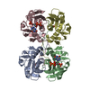 1a96C  1a97C  1nulS S: Starting model for refinement C: citing same article ( |
|---|---|
| Similar structure data |


-Links
 PDBj
PDBj
- Assembly
| Deposited unit | 
| ||||||||
|---|---|---|---|---|---|---|---|---|---|
| 1 | 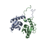
| ||||||||
| Unit cell |
|
-Components
| #1: Protein | Mass: 16959.502 Da / Num. of mol.: 2 / Mutation: C59A Source method: isolated from a genetically manipulated source Source: (gene. exp.) Escherichia coli (E. coli) / Strain: HB101 / Cellular location: CYTOPLASM / Gene: GPT / Plasmid: PT7-7 / Cellular location (production host): CYTOPLASM / Culture collection (production host): ATCC 87050 / Gene (production host): GPT / Production host: Escherichia coli (E. coli) / Strain (production host): SPHI606References: UniProt: P0A9M5, xanthine phosphoribosyltransferase#2: Water | ChemComp-HOH / | Water Mass: 18.015 Da / Num. of mol.: 66 / Source method: isolated from a natural source / Formula: H2O Mass: 18.015 Da / Num. of mol.: 66 / Source method: isolated from a natural source / Formula: H2O |
|---|
-Experimental details
-Experiment
| Experiment | Method: X-RAY DIFFRACTION / Number of used crystals: 1 |
|---|
- Sample preparation
| Crystal | Density Matthews: 2.15 Å3/Da / Density % sol: 43 % | ||||||||||||||||||||||||||||||||||||||||||
|---|---|---|---|---|---|---|---|---|---|---|---|---|---|---|---|---|---|---|---|---|---|---|---|---|---|---|---|---|---|---|---|---|---|---|---|---|---|---|---|---|---|---|---|
| Crystal grow | pH: 8 Details: XPRT WAS CRYSTALLIZED FROM 20% PEG4000 IN 0.1 M TRIS-HCL, pH 8.0 | ||||||||||||||||||||||||||||||||||||||||||
| Crystal grow | *PLUS pH: 7.6 / Method: vapor diffusion, hanging drop | ||||||||||||||||||||||||||||||||||||||||||
| Components of the solutions | *PLUS
|
-Data collection
| Diffraction | Mean temperature: 289 K |
|---|---|
| Diffraction source | Source: ROTATING ANODE / Type: RIGAKU RUH2R / Wavelength: 1.5418 |
| Detector | Type: RIGAKU RAXIS IIC / Detector: IMAGE PLATE / Date: Mar 29, 1994 / Details: MIRRORS |
| Radiation | Monochromator: NI FILTER / Monochromatic (M) / Laue (L): M / Scattering type: x-ray |
| Radiation wavelength | Wavelength: 1.5418 Å / Relative weight: 1 |
| Reflection | Resolution: 2.25→60 Å / Num. obs: 11089 / % possible obs: 79.5 % / Observed criterion σ(I): 0 / Redundancy: 1.5 % / Biso Wilson estimate: 24 Å2 / Rmerge(I) obs: 0.037 / Rsym value: 0.037 / Net I/σ(I): 11.8 |
| Reflection shell | Resolution: 2.25→2.33 Å / Redundancy: 1.4 % / Rmerge(I) obs: 0.172 / Mean I/σ(I) obs: 3.4 / Rsym value: 0.172 / % possible all: 66.9 |
| Reflection | *PLUS Lowest resolution: 50 Å / Num. measured all: 16902 / Rmerge(I) obs: 0.037 |
| Reflection shell | *PLUS % possible obs: 66.9 % / Rmerge(I) obs: 0.172 |
- Processing
| Software |
| ||||||||||||||||||||||||||||||||||||||||||||||||||||||||||||||||||||||||||||||||
|---|---|---|---|---|---|---|---|---|---|---|---|---|---|---|---|---|---|---|---|---|---|---|---|---|---|---|---|---|---|---|---|---|---|---|---|---|---|---|---|---|---|---|---|---|---|---|---|---|---|---|---|---|---|---|---|---|---|---|---|---|---|---|---|---|---|---|---|---|---|---|---|---|---|---|---|---|---|---|---|---|---|
| Refinement | Method to determine structure: MOLECULAR REPLACEMENT MOLECULAR REPLACEMENT Starting model: PDB ENTRY 1NUL Resolution: 2.25→50 Å / Rfactor Rfree error: 0.009 / Data cutoff high absF: 100000 / Data cutoff low absF: 1 / Isotropic thermal model: RESTRAINED / Cross valid method: THROUGHOUT / σ(F): 0 / Details: BULK SOLVENT MODEL USED
| ||||||||||||||||||||||||||||||||||||||||||||||||||||||||||||||||||||||||||||||||
| Displacement parameters | Biso mean: 36.1 Å2 | ||||||||||||||||||||||||||||||||||||||||||||||||||||||||||||||||||||||||||||||||
| Refine analyze |
| ||||||||||||||||||||||||||||||||||||||||||||||||||||||||||||||||||||||||||||||||
| Refinement step | Cycle: LAST / Resolution: 2.25→50 Å
| ||||||||||||||||||||||||||||||||||||||||||||||||||||||||||||||||||||||||||||||||
| Refine LS restraints |
| ||||||||||||||||||||||||||||||||||||||||||||||||||||||||||||||||||||||||||||||||
| LS refinement shell | Resolution: 2.25→2.39 Å / Rfactor Rfree error: 0.038 / Total num. of bins used: 6
| ||||||||||||||||||||||||||||||||||||||||||||||||||||||||||||||||||||||||||||||||
| Xplor file |
| ||||||||||||||||||||||||||||||||||||||||||||||||||||||||||||||||||||||||||||||||
| Refinement | *PLUS % reflection Rfree: 10 % / Rfactor obs: 0.21 / Rfactor Rfree: 0.224 / Rfactor Rwork: 0.21 | ||||||||||||||||||||||||||||||||||||||||||||||||||||||||||||||||||||||||||||||||
| Solvent computation | *PLUS | ||||||||||||||||||||||||||||||||||||||||||||||||||||||||||||||||||||||||||||||||
| Displacement parameters | *PLUS | ||||||||||||||||||||||||||||||||||||||||||||||||||||||||||||||||||||||||||||||||
| Refine LS restraints | *PLUS
| ||||||||||||||||||||||||||||||||||||||||||||||||||||||||||||||||||||||||||||||||
| LS refinement shell | *PLUS Rfactor Rfree: 0.326 / Rfactor Rwork: 0.308 |
