 Movie
Movie Controller


+ Open data

- Basic information
| Entry | Database: PDB / ID: 1a6j | ||||||
|---|---|---|---|---|---|---|---|
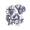
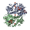
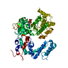
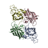
| Title | NITROGEN REGULATORY BACTERIAL PROTEIN IIA-NITROGEN | ||||||
 Components Components | NITROGEN REGULATORY IIA PROTEIN | ||||||
 Keywords Keywords | PHOSPHOTRANSFERASE SYSTEM / NITROGEN REGULATION | ||||||
| Function / homology |  Function and homology information Function and homology informationregulation of monoatomic ion transmembrane transporter activity / protein-N(PI)-phosphohistidine-sugar phosphotransferase activity / phosphoenolpyruvate-dependent sugar phosphotransferase system / enzyme inhibitor activity / protein kinase activator activity / : / kinase activity / cytosol Similarity search - Function | ||||||
| Biological species |  | ||||||
| Method |  X-RAY DIFFRACTION / MIR / Resolution: 2.35 Å X-RAY DIFFRACTION / MIR / Resolution: 2.35 Å | ||||||
 Authors Authors | Bordo, D. / Van Montfort, R. / Pijning, T. / Kalk, K.H. / Reizer, J. / Saier, M.H. / Dijkstra, B.W. | ||||||
 Citation Citation | Journal: J.Mol.Biol. / Year: 1998 Title: The three-dimensional structure of the nitrogen regulatory protein IIANtr from Escherichia coli. Authors: Bordo, D. / van Monfort, R.L. / Pijning, T. / Kalk, K.H. / Reizer, J. / Saier Jr., M.H. / Dijkstra, B.W. | ||||||
| History |
|
- Structure visualization
| Structure viewer | Molecule: MolmilJmol/JSmol |
|---|
- Downloads & links
-Download
| PDBx/mmCIF format | 1a6j.cif.gz | 71.5 KB | Display | PDBx/mmCIF format |
|---|---|---|---|---|
| PDB format | pdb1a6j.ent.gz | 54.5 KB | Display | PDB format |
| PDBx/mmJSON format | 1a6j.json.gz | Tree view | PDBx/mmJSON format | |
| Others |  Other downloads Other downloads |
-Validation report
| Summary document | 1a6j_validation.pdf.gz | 447.4 KB | Display | wwPDB validaton report |
|---|---|---|---|---|
| Full document | 1a6j_full_validation.pdf.gz | 451.1 KB | Display | |
| Data in XML | 1a6j_validation.xml.gz | 13.9 KB | Display | |
| Data in CIF | 1a6j_validation.cif.gz | 18.4 KB | Display | |
| Arichive directory | https://data.pdbj.org/pub/pdb/validation_reports/a6/1a6jftp://data.pdbj.org/pub/pdb/validation_reports/a6/1a6j | HTTPS FTP |
-Related structure data
| Similar structure data |
|---|


-Links
 PDBj
PDBj


- Assembly
| Deposited unit | 
| ||||||||
|---|---|---|---|---|---|---|---|---|---|
| 1 | 
| ||||||||
| Unit cell |
|
-Components
| #1: Protein | Mass: 17981.541 Da / Num. of mol.: 2 Source method: isolated from a genetically manipulated source Source: (gene. exp.) References: UniProt: P69829, protein-Npi-phosphohistidine-sugar phosphotransferase #2: Chemical |   Mass: 78.133 Da / Num. of mol.: 2 / Source method: obtained synthetically / Formula: C2H6OS Mass: 78.133 Da / Num. of mol.: 2 / Source method: obtained synthetically / Formula: C2H6OS#3: Chemical | ChemComp-SO4 / |   Mass: 96.063 Da / Num. of mol.: 1 / Source method: obtained synthetically / Formula: SO4 Mass: 96.063 Da / Num. of mol.: 1 / Source method: obtained synthetically / Formula: SO4#4: Water | ChemComp-HOH / |  Mass: 18.015 Da / Num. of mol.: 65 / Source method: isolated from a natural source / Formula: H2O Mass: 18.015 Da / Num. of mol.: 65 / Source method: isolated from a natural source / Formula: H2O |
|---|
-Experimental details
-Experiment
| Experiment | Method: X-RAY DIFFRACTION / Number of used crystals: 1 |
|---|
- Sample preparation
| Crystal | Density Matthews: 2.6 Å3/Da / Density % sol: 53 % | ||||||||||||||||||||||||||||||||||||||||||||||||||||||||||||
|---|---|---|---|---|---|---|---|---|---|---|---|---|---|---|---|---|---|---|---|---|---|---|---|---|---|---|---|---|---|---|---|---|---|---|---|---|---|---|---|---|---|---|---|---|---|---|---|---|---|---|---|---|---|---|---|---|---|---|---|---|---|
| Crystal grow | pH: 7.5 Details: PROTEIN CRYSTALLIZED WITH THE HANGING DROP SETUP. RESERVOIR CONTAINING 1.8 M AMMONIUM SULPHATE, 1 MM SODIUM AZIDE, 2MM DTT AND 0.1 M BES-NAOH, PH 7.5. | ||||||||||||||||||||||||||||||||||||||||||||||||||||||||||||
| Crystal | *PLUS Density % sol: 53 % | ||||||||||||||||||||||||||||||||||||||||||||||||||||||||||||
| Crystal grow | *PLUS Method: vapor diffusion, hanging drop | ||||||||||||||||||||||||||||||||||||||||||||||||||||||||||||
| Components of the solutions | *PLUS
|
-Data collection
| Diffraction | Mean temperature: 300 K |
|---|---|
| Diffraction source | Source: ROTATING ANODE / Type: ELLIOTT GX-21 / Wavelength: 1.5418 |
| Detector | Type: MACSCIENCE / Detector: IMAGE PLATE / Date: Jun 15, 1995 |
| Radiation | Monochromator: CU / Monochromatic (M) / Laue (L): M / Scattering type: x-ray |
| Radiation wavelength | Wavelength: 1.5418 Å / Relative weight: 1 |
| Reflection | Resolution: 2.35→30 Å / Num. obs: 14482 / % possible obs: 89 % / Observed criterion σ(I): 2 / Redundancy: 5.4 % / Biso Wilson estimate: 32 Å2 / Rmerge(I) obs: 0.05 / Rsym value: 0.06 / Net I/σ(I): 14 |
| Reflection shell | Resolution: 2.35→2.43 Å / Redundancy: 2.5 % / Rmerge(I) obs: 0.06 / Mean I/σ(I) obs: 5 / Rsym value: 0.2 / % possible all: 75 |
| Reflection | *PLUS Num. measured all: 104938 |
| Reflection shell | *PLUS % possible obs: 75 % / Rmerge(I) obs: 0.2 |
- Processing
| Software |
| ||||||||||||||||||||||||||||||||||||||||||||||||||||||||||||
|---|---|---|---|---|---|---|---|---|---|---|---|---|---|---|---|---|---|---|---|---|---|---|---|---|---|---|---|---|---|---|---|---|---|---|---|---|---|---|---|---|---|---|---|---|---|---|---|---|---|---|---|---|---|---|---|---|---|---|---|---|---|
| Refinement | Method to determine structure: MIR / Resolution: 2.35→40 Å / Rfactor Rfree error: 0.01 / Data cutoff high absF: 10000000 / Data cutoff low absF: 0 / Isotropic thermal model: 0 / Cross valid method: THROUGHOUT / σ(F): 0
| ||||||||||||||||||||||||||||||||||||||||||||||||||||||||||||
| Displacement parameters | Biso mean: 24 Å2 | ||||||||||||||||||||||||||||||||||||||||||||||||||||||||||||
| Refine analyze | Luzzati coordinate error obs: 0.24 Å / Luzzati d res low obs: 5 Å / Luzzati sigma a obs: 0.22 Å | ||||||||||||||||||||||||||||||||||||||||||||||||||||||||||||
| Refinement step | Cycle: LAST / Resolution: 2.35→40 Å
| ||||||||||||||||||||||||||||||||||||||||||||||||||||||||||||
| Refine LS restraints |
| ||||||||||||||||||||||||||||||||||||||||||||||||||||||||||||
| LS refinement shell | Resolution: 2.35→2.46 Å / Rfactor Rfree error: 0.042 / Total num. of bins used: 8
| ||||||||||||||||||||||||||||||||||||||||||||||||||||||||||||
| Xplor file |
|
