Movie
Movie Controller


+ Open data

- Basic information
| Entry | Database: EMDB / ID: EMD-9901 | ||||||||||||
|---|---|---|---|---|---|---|---|---|---|---|---|---|---|
| Title | Helical Reconstruction of S-OPA1 At Nucleotide-free State | ||||||||||||
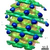
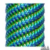
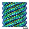
 Map data Map data | This is the map by helical reconstruction using IHRSR for SOPA-1 of the nucleotide free state. The resolution is about 15 angstrom by FSC 0.143 criterion. | ||||||||||||
 Sample Sample |
| ||||||||||||
| Biological species |   Homo sapiens (human) Homo sapiens (human) | ||||||||||||
| Method | helical reconstruction / cryo EM / Resolution: 15.0 Å | ||||||||||||
 Authors Authors | Zhang D / Zhang Y / Sun F | ||||||||||||
| Funding support |  China, 3 items China, 3 items
| ||||||||||||
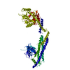
 Citation Citation | Journal: Elife / Year: 2020 Title: Cryo-EM structures of S-OPA1 reveal its interactions with membrane and changes upon nucleotide binding. Authors: Danyang Zhang / Yan Zhang / Jun Ma / Chunmei Zhu / Tongxin Niu / Wenbo Chen / Xiaoyun Pang / Yujia Zhai / Fei Sun / Abstract: Mammalian mitochondrial inner membrane fusion is mediated by optic atrophy 1 (OPA1). Under physiological conditions, OPA1 undergoes proteolytic processing to form a membrane-anchored long isoform (L- ...Mammalian mitochondrial inner membrane fusion is mediated by optic atrophy 1 (OPA1). Under physiological conditions, OPA1 undergoes proteolytic processing to form a membrane-anchored long isoform (L-OPA1) and a soluble short isoform (S-OPA1). A combination of L-OPA1 and S-OPA1 is essential for efficient membrane fusion; however, the relevant mechanism is not well understood. In this study, we investigate the cryo-electron microscopic structures of S-OPA1-coated liposomes in nucleotide-free and GTPγS-bound states. S-OPA1 exhibits a general dynamin-like structure and can assemble onto membranes in a helical array with a dimer building block. We reveal that hydrophobic residues in its extended membrane-binding domain are critical for its tubulation activity. The binding of GTPγS triggers a conformational change and results in a rearrangement of the helical lattice and tube expansion similar to that of S-Mgm1. These observations indicate that S-OPA1 adopts a dynamin-like power stroke membrane remodeling mechanism during mitochondrial inner membrane fusion. | ||||||||||||
| History |
|
- Structure visualization
| Movie |
 Movie viewer Movie viewer |
|---|---|
| Structure viewer | EM map: SurfViewMolmilJmol/JSmol |
| Supplemental images |

- Downloads & links
-EMDB archive
| Map data | emd_9901.map.gz | 197 MB | EMDB map data format | |
|---|---|---|---|---|
| Header (meta data) | emd-9901-v30.xmlemd-9901.xml | 16.3 KB 16.3 KB | Display Display | EMDB header |
| FSC (resolution estimation) | emd_9901_fsc.xml | 14.1 KB | Display | FSC data file |
| Images | 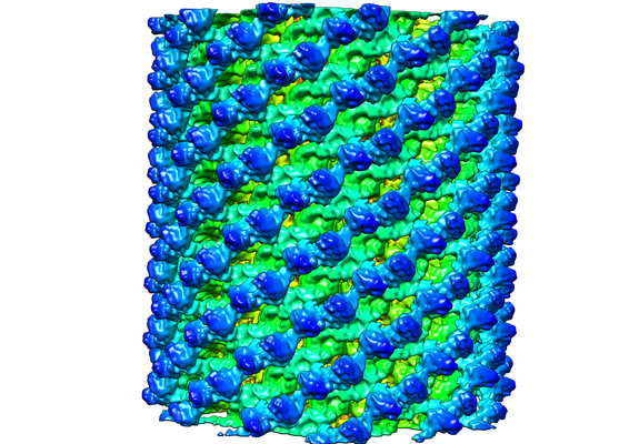 emd_9901.png emd_9901.png | 286.7 KB | ||
| Archive directory |  http://ftp.pdbj.org/pub/emdb/structures/EMD-9901ftp://ftp.pdbj.org/pub/emdb/structures/EMD-9901 http://ftp.pdbj.org/pub/emdb/structures/EMD-9901ftp://ftp.pdbj.org/pub/emdb/structures/EMD-9901 | HTTPS FTP |
-Related structure data


-Links
| EMDB pages | EMDB (EBI/PDBe) / EMDataResource |
|---|
-Map
| File | Download / File: emd_9901.map.gz / Format: CCP4 / Size: 216 MB / Type: IMAGE STORED AS FLOATING POINT NUMBER (4 BYTES) | ||||||||||||||||||||||||||||||||||||||||||||||||||||||||||||||||||||
|---|---|---|---|---|---|---|---|---|---|---|---|---|---|---|---|---|---|---|---|---|---|---|---|---|---|---|---|---|---|---|---|---|---|---|---|---|---|---|---|---|---|---|---|---|---|---|---|---|---|---|---|---|---|---|---|---|---|---|---|---|---|---|---|---|---|---|---|---|---|
| Annotation | This is the map by helical reconstruction using IHRSR for SOPA-1 of the nucleotide free state. The resolution is about 15 angstrom by FSC 0.143 criterion. | ||||||||||||||||||||||||||||||||||||||||||||||||||||||||||||||||||||
| Voxel size | X=Y=Z: 1.42 Å | ||||||||||||||||||||||||||||||||||||||||||||||||||||||||||||||||||||
| Density |
| ||||||||||||||||||||||||||||||||||||||||||||||||||||||||||||||||||||
| Symmetry | Space group: 1 | ||||||||||||||||||||||||||||||||||||||||||||||||||||||||||||||||||||
| Details | EMDB XML:
CCP4 map header:
| ||||||||||||||||||||||||||||||||||||||||||||||||||||||||||||||||||||
-Supplemental data
- Sample components
-Entire : truncated S-OPA1(253-960) coated liposomal tube
| Entire | Name: truncated S-OPA1(253-960) coated liposomal tube |
|---|---|
| Components |
|
-Supramolecule #1: truncated S-OPA1(253-960) coated liposomal tube
| Supramolecule | Name: truncated S-OPA1(253-960) coated liposomal tube / type: complex / ID: 1 / Parent: 0 / Macromolecule list: all Details: 1mg/ml S-OPA1(253-960) was incubated with 1mg/ml liposome at room temperature for 30 min. |
|---|---|
| Source (natural) | Organism: Homo sapiens (human) / Organelle: Mitochondrion / Location in cell: Mitochondrion inner membrane |
| Recombinant expression | Organism:  Escherichia coli (E. coli) / Recombinant strain: Rosetta (DE3) / Recombinant plasmid: pET32M-3C Escherichia coli (E. coli) / Recombinant strain: Rosetta (DE3) / Recombinant plasmid: pET32M-3C |
-Macromolecule #1: Dynamin-like 120 kDa protein, mitochondrial, short form for isofo...
| Macromolecule | Name: Dynamin-like 120 kDa protein, mitochondrial, short form for isoform 1; Optic atrophy protein 1 (OPA1), short form for isoform 1 type: protein_or_peptide / ID: 1 / Enantiomer: LEVO |
|---|---|
| Source (natural) | Organism: Homo sapiens (human) |
| Recombinant expression | Organism: Escherichia coli (E. coli) |
| Sequence | String: GPGSDKGIHH RKLKKSLIDM YSEVLDVLSD YDASYNTQDH LPRVVVVGDQ SAGKTSVLEM IAQARIFPRG SGEMMTRSPV KVTLSEGPHH VALFKDSSRE FDLTKEEDLA ALRHEIELRM RKNVKEGCTV SPETISLNVK GPGLQRMVLV DLPGVINTVT SGMAPDTKET ...String: GPGSDKGIHH RKLKKSLIDM YSEVLDVLSD YDASYNTQDH LPRVVVVGDQ SAGKTSVLEM IAQARIFPRG SGEMMTRSPV KVTLSEGPHH VALFKDSSRE FDLTKEEDLA ALRHEIELRM RKNVKEGCTV SPETISLNVK GPGLQRMVLV DLPGVINTVT SGMAPDTKET IFSISKAYMQ NPNAIILCIQ DGSVDAERSI VTDLVSQMDP HGRRTIFVLT KVDLAEKNVA SPSRIQQIIE GKLFPMKALG YFAVVTGKGN SSESIEAIRE YEEEFFQNSK LLKTSMLKAH QVTTRNLSLA VSDCFWKMVR ESVEQQADSF KATRFNLETE WKNNYPRLRE LDRNELFEKA KNEILDEVIS LSQVTPKHWE EILQQSLWER VSTHVIENIY LPAAQTMNSG TFNTTVDIKL KQWTDKQLPN KAVEVAWETL QEEFSRFMTE PKGKEHDDIF DKLKEAVKEE SIKRHKWNDF AEDSLRVIQH NALEDRSISD KQQWDAAIYF MEEALQARLK DTENAIENMV GPDWKKRWLY WKNRTQEQCV HNETKNELEK MLKCNEEHPA YLASDEITTV RKNLESRGVE VDPSLIKDTW HQVYRRHFLK TALNHCNLCR RGFYYYQRHF VDSELECNDV VLFWRIQRML AITANTLRQQ LTNTEVRRLE KNVKEVLEDF AEDGEKKIKL LTGKRVQLAE DLKKVREIQE KLDAFIEALH QEK |
-Experimental details
-Structure determination
| Method | cryo EM |
|---|---|
 Processing Processing | helical reconstruction |
| Aggregation state | helical array |
-Sample preparation
| Concentration | 1 mg/mL | ||||||||||||
|---|---|---|---|---|---|---|---|---|---|---|---|---|---|
| Buffer | pH: 8 Component:
| ||||||||||||
| Grid | Model: Quantifoil R2/1 / Material: COPPER / Mesh: 300 / Support film - Material: CARBON / Support film - topology: HOLEY ARRAY / Pretreatment - Type: GLOW DISCHARGE / Pretreatment - Atmosphere: OTHER | ||||||||||||
| Vitrification | Cryogen name: ETHANE / Chamber humidity: 100 % / Chamber temperature: 289 K / Instrument: FEI VITROBOT MARK IV Details: The grid was blotted 3s with a force 1 before plunging. |
- Electron microscopy
| Microscope | FEI TITAN KRIOS |
|---|---|
| Electron beam | Acceleration voltage: 300 kV / Electron source: FIELD EMISSION GUN |
| Electron optics | C2 aperture diameter: 50.0 µm / Calibrated magnification: 98591 / Illumination mode: FLOOD BEAM / Imaging mode: BRIGHT FIELDBright-field microscopy / Cs: 2.7 mm / Nominal defocus max: 4.0 µm / Nominal defocus min: 2.0 µm / Nominal magnification: 59000 |
| Sample stage | Specimen holder model: FEI TITAN KRIOS AUTOGRID HOLDER / Cooling holder cryogen: NITROGEN |
| Temperature | Min: 77.0 K / Max: 82.0 K |
| Image recording | Film or detector model: FEI FALCON III (4k x 4k) / Detector mode: INTEGRATING / Digitization - Dimensions - Width: 4096 pixel / Digitization - Dimensions - Height: 4096 pixel / Digitization - Sampling interval: 14.0 µm / Number real images: 2112 / Average exposure time: 2.0 sec. / Average electron dose: 25.0 e/Å2 |
| Experimental equipment |  Model: Titan Krios / Image courtesy: FEI Company |
-Image processing
| Segment selection | Number selected: 6644 / Software - Name: SPIDER (ver. spiderweb.24.08) Software - details: a spider script with operation command wi was used to cut the tubes into segments Details: Tubes were boxed out using the tool e2helixboxer.py of EMAN2. Diameter classification were performed for all the boxed tubes. Then 6644 segments of particles were cutting out with an overlap ...Details: Tubes were boxed out using the tool e2helixboxer.py of EMAN2. Diameter classification were performed for all the boxed tubes. Then 6644 segments of particles were cutting out with an overlap of 94% for the main diameter class 53nm. |
|---|---|
| CTF correction | Software - Name: Gctf (ver. 1.18) |
| Startup model | Type of model: OTHER Details: A cylinder was generated by SPIDER with diameter same as the main diameter class 53nm of S-OPA1 and used as starting model to avoid model bias. |
| Final angle assignment | Type: NOT APPLICABLE / Software - Name: SPIDER (ver. spiderweb.24.08) Details: Final angles were determined by projection matching calculation. |
| Final reconstruction | Number classes used: 1 Applied symmetry - Helical parameters - Δz: 27.0 Å Applied symmetry - Helical parameters - Δ&Phi: 20.87 ° Applied symmetry - Helical parameters - Axial symmetry: C6 (6 fold cyclic )Algorithm: BACK PROJECTION / Resolution.type: BY AUTHOR / Resolution: 15.0 Å / Resolution method: FSC 0.143 CUT-OFF / Software - Name: SPIDER (ver. spiderweb.24.08) / Number images used: 5437 |
| FSC plot (resolution estimation) |  |
-Atomic model buiding 1
| Initial model | PDB ID: Chain - Chain ID: A |
|---|---|
| Refinement | Space: REAL / Protocol: RIGID BODY FIT / Overall B value: 1000 / Target criteria: Cross correlation |