 Movie
Movie Controller


+ Open data

- Basic information
| Entry | Database: EMDB / ID: EMD-9796 | |||||||||
|---|---|---|---|---|---|---|---|---|---|---|
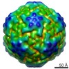
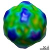
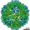
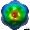
| Title | AAV5 in complex with AAVR | |||||||||
 Map data Map data | ||||||||||
 Sample Sample |
| |||||||||
 Keywords Keywords |  adeno-associated virus / AAV5 / receptor / AAVR / VIRUS adeno-associated virus / AAV5 / receptor / AAVR / VIRUS | |||||||||
| Function / homology |  Function and homology information Function and homology informationT=1 icosahedral viral capsid / neuron migration / cytoplasmic vesicle / Golgi membrane / nucleolus / structural molecule activity / Golgi apparatus / membrane / plasma membraneSimilarity search - Function | |||||||||
| Biological species |  Adeno-associated virus - 5 / Adeno-associated virus - 5 /  Homo sapiens (human) Homo sapiens (human) | |||||||||
| Method | single particle reconstruction / cryo EM / Resolution: 3.18 Å | |||||||||
 Authors Authors | Lou Z / Zhang R | |||||||||
| Funding support |  China, 1 items China, 1 items
| |||||||||
 Citation Citation | Journal: Nat Commun / Year: 2019 Title: Divergent engagements between adeno-associated viruses with their cellular receptor AAVR. Authors: Ran Zhang / Guangxue Xu / Lin Cao / Zixian Sun / Yong He / Mengtian Cui / Yuna Sun / Shentao Li / Huapeng Li / Lan Qin / Mingxu Hu / Zhengjia Yuan / Zipei Rao / Wei Ding / Zihe Rao / Zhiyong Lou / Abstract: Adeno-associated virus (AAV) receptor (AAVR) is an essential receptor for the entry of multiple AAV serotypes with divergent rules; however, the mechanism remains unclear. Here, we determine the ...Adeno-associated virus (AAV) receptor (AAVR) is an essential receptor for the entry of multiple AAV serotypes with divergent rules; however, the mechanism remains unclear. Here, we determine the structures of the AAV1-AAVR and AAV5-AAVR complexes, revealing the molecular details by which PKD1 recognizes AAV5 and PKD2 is solely engaged with AAV1. PKD2 lies on the plateau region of the AAV1 capsid. However, the AAV5-AAVR interface is strikingly different, in which PKD1 is bound at the opposite side of the spike of the AAV5 capsid than the PKD2-interacting region of AAV1. Residues in strands F/G and the CD loop of PKD1 interact directly with AAV5, whereas residues in strands B/C/E and the BC loop of PKD2 make contact with AAV1. These findings further the understanding of the distinct mechanisms by which AAVR recognizes various AAV serotypes and provide an example of a single receptor engaging multiple viral serotypes with divergent rules. | |||||||||
| History |
|
- Structure visualization
| Movie |
Movie viewer |
|---|---|
| Structure viewer | EM map: SurfViewMolmilJmol/JSmol |
| Supplemental images |

- Downloads & links
-EMDB archive
| Map data | emd_9796.map.gz | 64.4 MB | EMDB map data format | |
|---|---|---|---|---|
| Header (meta data) | emd-9796-v30.xmlemd-9796.xml | 11.3 KB 11.3 KB | Display Display | EMDB header |
| Images | 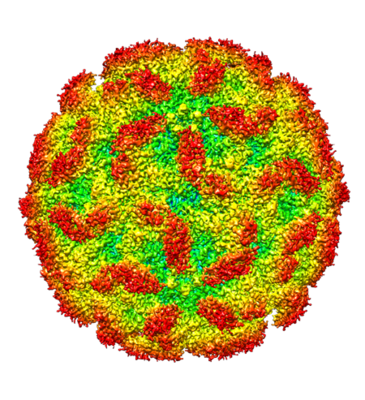 emd_9796.png emd_9796.png | 241.2 KB | ||
| Filedesc metadata | emd-9796.cif.gz | 5.5 KB | ||
| Archive directory |  http://ftp.pdbj.org/pub/emdb/structures/EMD-9796ftp://ftp.pdbj.org/pub/emdb/structures/EMD-9796 http://ftp.pdbj.org/pub/emdb/structures/EMD-9796ftp://ftp.pdbj.org/pub/emdb/structures/EMD-9796 | HTTPS FTP |
-Related structure data
| Related structure data |  6jcsMC  9794C  9795C 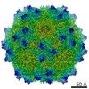 9797C 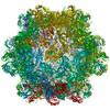 6jcqC 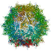 6jcrC 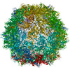 6jctC M: atomic model generated by this map C: citing same article ( |
|---|---|
| Similar structure data |

-Links
| EMDB pages | EMDB (EBI/PDBe) / EMDataResource |
|---|---|
| Related items in Molecule of the Month |


-Map
| File | Download / File: emd_9796.map.gz / Format: CCP4 / Size: 209.3 MB / Type: IMAGE STORED AS FLOATING POINT NUMBER (4 BYTES) | ||||||||||||||||||||||||||||||||||||||||||||||||||||||||||||||||||||
|---|---|---|---|---|---|---|---|---|---|---|---|---|---|---|---|---|---|---|---|---|---|---|---|---|---|---|---|---|---|---|---|---|---|---|---|---|---|---|---|---|---|---|---|---|---|---|---|---|---|---|---|---|---|---|---|---|---|---|---|---|---|---|---|---|---|---|---|---|---|
| Voxel size | X=Y=Z: 0.93 Å | ||||||||||||||||||||||||||||||||||||||||||||||||||||||||||||||||||||
| Density |
| ||||||||||||||||||||||||||||||||||||||||||||||||||||||||||||||||||||
| Symmetry | Space group: 1 | ||||||||||||||||||||||||||||||||||||||||||||||||||||||||||||||||||||
| Details | EMDB XML:
CCP4 map header:
| ||||||||||||||||||||||||||||||||||||||||||||||||||||||||||||||||||||
-Supplemental data
- Sample components
-Entire : Adeno-associated virus - 5
| Entire | Name: Adeno-associated virus - 5 |
|---|---|
| Components |
|
-Supramolecule #1: Adeno-associated virus - 5
| Supramolecule | Name: Adeno-associated virus - 5 / type: complex / ID: 1 / Parent: 0 / Macromolecule list: all |
|---|
-Supramolecule #2: capsid protein VP1
| Supramolecule | Name: capsid protein VP1 / type: complex / ID: 2 / Parent: 1 / Macromolecule list: #1 |
|---|---|
| Source (natural) | Organism: Adeno-associated virus - 5 |
-Supramolecule #3: PKD1
| Supramolecule | Name: PKD1 / type: organelle_or_cellular_component / ID: 3 / Parent: 1 / Macromolecule list: #2 |
|---|---|
| Source (natural) | Organism: Homo sapiens (human) |
-Macromolecule #1: Capsid protein
| Macromolecule | Name: Capsid protein / type: protein_or_peptide / ID: 1 / Number of copies: 1 / Enantiomer: LEVO |
|---|---|
| Source (natural) | Organism: Adeno-associated virus - 5 |
| Molecular weight | Theoretical: 58.213078 KDa |
| Sequence | String: DGVGNASGDW HCDSTWMGDR VVTKSTRTWV LPSYNNHQYR EIKSGSVDGS NANAYFGYST PWGYFDFNRF HSHWSPRDWQ RLINNYWGF RPRSLRVKIF NIQVKEVTVQ DSTTTIANNL TSTVQVFTDD DYQLPYVVGN GTEGCLPAFP PQVFTLPQYG Y ATLNRDNT ...String: DGVGNASGDW HCDSTWMGDR VVTKSTRTWV LPSYNNHQYR EIKSGSVDGS NANAYFGYST PWGYFDFNRF HSHWSPRDWQ RLINNYWGF RPRSLRVKIF NIQVKEVTVQ DSTTTIANNL TSTVQVFTDD DYQLPYVVGN GTEGCLPAFP PQVFTLPQYG Y ATLNRDNT ENPTERSSFF CLEYFPSKML RTGNNFEFTY NFEEVPFHSS FAPSQNLFKL ANPLVDQYLY RFVSTNNTGG VQ FNKNLAG RYANTYKNWF PGPMGRTQGW NLGSGVNRAS VSAFATTNRM ELEGASYQVP PQPNGMTNNL QGSNTYALEN TMI FNSQPA NPGTTATYLE GNMLITSESE TQPVNRVAYN VGGQMATNNQ SSTTAPATGT YNLQEIVPGS VWMERDVYLQ GPIW AKIPE TGAHFHPSPA MGGFGLKHPP PMMLIKNTPV PGNITSFSDV PVSSFITQYS TGQVTVEMEW ELKKENSKRW NPEIQ YTNN YNDPQFVDFA PDSTGEYRTT RPIGTRYLTR PL UniProtKB: Capsid protein |
-Macromolecule #2: Dyslexia-associated protein KIAA0319-like protein
| Macromolecule | Name: Dyslexia-associated protein KIAA0319-like protein / type: protein_or_peptide / ID: 2 / Number of copies: 1 / Enantiomer: LEVO |
|---|---|
| Source (natural) | Organism: Homo sapiens (human) |
| Molecular weight | Theoretical: 10.911322 KDa |
| Recombinant expression | Organism:  Escherichia coli (E. coli) Escherichia coli (E. coli) |
| Sequence | String: VIKELVVSAG ESVQITLPKN EVQLNAYVLQ EPPKGETYTY DWQLITHPRD YSGEMEGKHS QILKLSKLTP GLYEFKVIVE GQNAHGEGY VNVTVKPE UniProtKB: Dyslexia-associated protein KIAA0319-like protein |
-Experimental details
-Structure determination
| Method | cryo EM |
|---|---|
 Processing Processing | single particle reconstruction |
| Aggregation state | 2D array |
-Sample preparation
| Buffer | pH: 8 |
|---|---|
| Vitrification | Cryogen name: ETHANE |
- Electron microscopy
| Microscope | FEI TECNAI ARCTICA |
|---|---|
| Electron beam | Acceleration voltage: 200 kV / Electron source: FIELD EMISSION GUN |
| Electron optics | Illumination mode: FLOOD BEAM / Imaging mode: BRIGHT FIELDBright-field microscopy |
| Image recording | Film or detector model: FEI FALCON II (4k x 4k) / Average electron dose: 36.0 e/Å2 |
| Experimental equipment |  Model: Talos Arctica / Image courtesy: FEI Company |
-Image processing
| Startup model | Type of model: PDB ENTRY |
|---|---|
| Initial angle assignment | Type: ANGULAR RECONSTITUTION |
| Final angle assignment | Type: ANGULAR RECONSTITUTION |
| Final reconstruction | Resolution.type: BY AUTHOR / Resolution: 3.18 Å / Resolution method: FSC 0.143 CUT-OFF / Number images used: 12590 |
