 Movie
Movie Controller


+ Open data

- Basic information
| Entry | Database: EMDB / ID: EMD-8403 | |||||||||
|---|---|---|---|---|---|---|---|---|---|---|
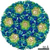
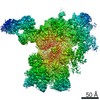
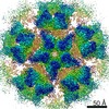
| Title | sub-tomogram average of in vitro assembled HIV-1 Gag VLPs | |||||||||
 Map data Map data | sub-Tomogram average of in vitro reconstituted HIV-1 virus-like particles imaged prior to protease treatement confirming immature conformation of the Gag lattice. | |||||||||
 Sample Sample |
| |||||||||
| Biological species |   Human immunodeficiency virus 1 Human immunodeficiency virus 1 | |||||||||
| Method | subtomogram averaging / cryo EM / Resolution: 18.0 Å | |||||||||
 Authors Authors | Himes BA / Zhang P | |||||||||
| Funding support |  United States, 1 items United States, 1 items
| |||||||||
 Citation Citation | Journal: Nat Commun / Year: 2016 Title: In vitro protease cleavage and computer simulations reveal the HIV-1 capsid maturation pathway. Authors: Jiying Ning / Gonca Erdemci-Tandogan / Ernest L Yufenyuy / Jef Wagner / Benjamin A Himes / Gongpu Zhao / Christopher Aiken / Roya Zandi / Peijun Zhang /  Abstract: HIV-1 virions assemble as immature particles containing Gag polyproteins that are processed by the viral protease into individual components, resulting in the formation of mature infectious particles. ...HIV-1 virions assemble as immature particles containing Gag polyproteins that are processed by the viral protease into individual components, resulting in the formation of mature infectious particles. There are two competing models for the process of forming the mature HIV-1 core: the disassembly and de novo reassembly model and the non-diffusional displacive model. To study the maturation pathway, we simulate HIV-1 maturation in vitro by digesting immature particles and assembled virus-like particles with recombinant HIV-1 protease and monitor the process with biochemical assays and cryoEM structural analysis in parallel. Processing of Gag in vitro is accurate and efficient and results in both soluble capsid protein and conical or tubular capsid assemblies, seemingly converted from immature Gag particles. Computer simulations further reveal probable assembly pathways of HIV-1 capsid formation. Combining the experimental data and computer simulations, our results suggest a sequential combination of both displacive and disassembly/reassembly processes for HIV-1 maturation. | |||||||||
| History |
|
- Structure visualization
| Movie |
 Movie viewer Movie viewer |
|---|---|
| Structure viewer | EM map: SurfViewMolmilJmol/JSmol |
| Supplemental images |

- Downloads & links
-EMDB archive
| Map data | emd_8403.map.gz | 891.1 KB | EMDB map data format | |
|---|---|---|---|---|
| Header (meta data) | emd-8403-v30.xmlemd-8403.xml | 16.4 KB 16.4 KB | Display Display | EMDB header |
| Images | 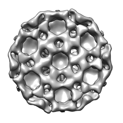 emd_8403.png emd_8403.png | 79.2 KB | ||
| Others | emd_8403_half_map_1.map.gzemd_8403_half_map_2.map.gz | 690.5 KB 711.4 KB | ||
| Archive directory |  http://ftp.pdbj.org/pub/emdb/structures/EMD-8403ftp://ftp.pdbj.org/pub/emdb/structures/EMD-8403 http://ftp.pdbj.org/pub/emdb/structures/EMD-8403ftp://ftp.pdbj.org/pub/emdb/structures/EMD-8403 | HTTPS FTP |
-Validation report
| Summary document | emd_8403_validation.pdf.gz | 386.8 KB | Display | EMDB validaton report |
|---|---|---|---|---|
| Full document | emd_8403_full_validation.pdf.gz | 386.4 KB | Display | |
| Data in XML | emd_8403_validation.xml.gz | 7.2 KB | Display | |
| Arichive directory | https://ftp.pdbj.org/pub/emdb/validation_reports/EMD-8403ftp://ftp.pdbj.org/pub/emdb/validation_reports/EMD-8403 | HTTPS FTP |
-Related structure data

-Links
| EMDB pages | EMDB (EBI/PDBe) / EMDataResource |
|---|
-Map
| File | Download / File: emd_8403.map.gz / Format: CCP4 / Size: 6.6 MB / Type: IMAGE STORED AS FLOATING POINT NUMBER (4 BYTES) | ||||||||||||||||||||||||||||||||||||||||||||||||||||||||||||||||||||
|---|---|---|---|---|---|---|---|---|---|---|---|---|---|---|---|---|---|---|---|---|---|---|---|---|---|---|---|---|---|---|---|---|---|---|---|---|---|---|---|---|---|---|---|---|---|---|---|---|---|---|---|---|---|---|---|---|---|---|---|---|---|---|---|---|---|---|---|---|---|
| Annotation | sub-Tomogram average of in vitro reconstituted HIV-1 virus-like particles imaged prior to protease treatement confirming immature conformation of the Gag lattice. | ||||||||||||||||||||||||||||||||||||||||||||||||||||||||||||||||||||
| Voxel size | X=Y=Z: 3.62 Å | ||||||||||||||||||||||||||||||||||||||||||||||||||||||||||||||||||||
| Density |
| ||||||||||||||||||||||||||||||||||||||||||||||||||||||||||||||||||||
| Symmetry | Space group: 1 | ||||||||||||||||||||||||||||||||||||||||||||||||||||||||||||||||||||
| Details | EMDB XML:
CCP4 map header:
| ||||||||||||||||||||||||||||||||||||||||||||||||||||||||||||||||||||
-Supplemental data
-Half map: gold-standard half map used for FSC calculation, masked...
| File | emd_8403_half_map_1.map | ||||||||||||
|---|---|---|---|---|---|---|---|---|---|---|---|---|---|
| Annotation | gold-standard half map used for FSC calculation, masked by soft cylinder. | ||||||||||||
| Projections & Slices |
| ||||||||||||
| Density Histograms |
 Z
Z Y
Y X
X


-Half map: gold-standard half map used for FSC calculation, masked...
| File | emd_8403_half_map_2.map | ||||||||||||
|---|---|---|---|---|---|---|---|---|---|---|---|---|---|
| Annotation | gold-standard half map used for FSC calculation, masked by soft cylinder. | ||||||||||||
| Projections & Slices |
| ||||||||||||
| Density Histograms |


- Sample components
-Entire : gag polyprotein, Pr55gag ([delta]-MA15-100[delta]p6)
| Entire | Name: gag polyprotein, Pr55gag ([delta]-MA15-100[delta]p6) |
|---|---|
| Components |
|
-Supramolecule #1: gag polyprotein, Pr55gag ([delta]-MA15-100[delta]p6)
| Supramolecule | Name: gag polyprotein, Pr55gag ([delta]-MA15-100[delta]p6) / type: complex / ID: 1 / Parent: 0 Details: PRR-55 construct, expressed recombinantly without protease. |
|---|---|
| Source (natural) | Organism: Human immunodeficiency virus 1 |
| Recombinant expression | Organism:  |
| Molecular weight | Theoretical: 39.9 KDa |
-Macromolecule #1: HIV-1 Gag VLPs
| Macromolecule | Name: HIV-1 Gag VLPs / type: protein_or_peptide / ID: 1 / Enantiomer: LEVO |
|---|---|
| Sequence | String: GARASVLSGG ELDRALDKIE EEQNKSKKKA QQAAADTGHS SQVSQNYPIV QNIQGQMVHQ AISPRTLNAW VKVVEEKAFS PEVIPMFSAL SEGATPQDLN TMLNTVGGHQ AAMQMLKETI NEEAAEWDRV HPVHAGPIAP GQMREPRGSD IAGTTSTLQE QIGWMTNNPP ...String: GARASVLSGG ELDRALDKIE EEQNKSKKKA QQAAADTGHS SQVSQNYPIV QNIQGQMVHQ AISPRTLNAW VKVVEEKAFS PEVIPMFSAL SEGATPQDLN TMLNTVGGHQ AAMQMLKETI NEEAAEWDRV HPVHAGPIAP GQMREPRGSD IAGTTSTLQE QIGWMTNNPP IPVGEIYKRW IILGLNKIVR MYSPTSILDI RQGPKEPFRD YVDRFYKTLR AEQASQEVKN WMTETLLVQN ANPDCKTILK ALGPAATLEE MMTACQGVGG PGHKARVLAE AMSQVTNTAT IMMQRGNFRN QRKMVKCFNC GKEGHTARNC RAPRKKGCWK CGKEGHQMKD CTERQANFLG KIWPSYKGRP |
-Experimental details
-Structure determination
| Method | cryo EM |
|---|---|
 Processing Processing | subtomogram averaging |
| Aggregation state | 3D array |
-Sample preparation
| Buffer | pH: 6 Details: 50 mM sodium acetate (pH6.0), 100 microM ZnSO4 and 5 mM DTT |
|---|---|
| Grid | Model: Quantifoil R2/2 / Support film - Material: CARBON / Support film - topology: HOLEY / Pretreatment - Type: GLOW DISCHARGE |
| Vitrification | Cryogen name: ETHANE / Chamber humidity: 100 % / Instrument: HOMEMADE PLUNGER |
| Details | The sample forms a 3-dimensional lattice with ~8nm spacing. The sub-tomogram averages are aligned along the c6 symmetry axis, such that ~ 12 assymetric units are included for a total effective mass of ~ 0.45 MDa |
- Electron microscopy
| Microscope | FEI POLARA 300 |
|---|---|
| Image recording | Film or detector model: FEI FALCON II (4k x 4k) / Detector mode: INTEGRATING / Digitization - Dimensions - Width: 4096 pixel / Digitization - Dimensions - Height: 4096 pixel / Digitization - Sampling interval: 14.0 µm / Average electron dose: 1.0 e/Å2 |
| Electron beam | Acceleration voltage: 300 kV / Electron source:  FIELD EMISSION GUN FIELD EMISSION GUN |
| Electron optics | Calibrated magnification: 77348 / Illumination mode: SPOT SCAN / Imaging mode: BRIGHT FIELD / Cs: 2.0 mm / Nominal defocus max: 5.0 µm / Nominal defocus min: 4.0 µm / Nominal magnification: 59000 |
| Experimental equipment |  Model: Tecnai Polara / Image courtesy: FEI Company |
-Image processing
| Final reconstruction | Applied symmetry - Point group: C6 (6 fold cyclic) / Algorithm: EXACT BACK PROJECTION / Resolution.type: BY AUTHOR / Resolution: 18.0 Å / Resolution method: FSC 0.143 CUT-OFF / Software - Name: matlab scripts / Number subtomograms used: 4000 |
|---|---|
| Extraction | Number tomograms: 28 / Number images used: 8400 / Reference model: EMD-2706 / Method: Cross-correlation based template matching / Software - Name: matlab scripts |
| CTF correction | Software - Name: matlab scripts Details: Phase flipping on the projections, amplitude correction applied to final average. |
| Final angle assignment | Type: NOT APPLICABLE |
