 Movie
Movie Controller


+ Open data

- Basic information
| Entry | Database: EMDB / ID: EMD-8365 | |||||||||
|---|---|---|---|---|---|---|---|---|---|---|
| Title | 10 nm RNA nanoprism | |||||||||
 Map data Map data | 10 nm RNA nanoprism | |||||||||
 Sample Sample |
| |||||||||
| Biological species | synthetic construct (others) | |||||||||
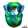
| Method |  single particle reconstruction / cryo EM / Resolution: 25.0 Å single particle reconstruction / cryo EM / Resolution: 25.0 Å | |||||||||
 Authors Authors | Khisamutdinov E / Jasinski D / Li H / Zhang K / Chiu W / Guo P | |||||||||
| Funding support |  United States, 2 items United States, 2 items
| |||||||||
 Citation Citation | Journal: Adv Mater / Year: 2016 Title: Fabrication of RNA 3D Nanoprisms for Loading and Protection of Small RNAs and Model Drugs. Authors: Emil F Khisamutdinov / Daniel L Jasinski / Hui Li / Kaiming Zhang / Wah Chiu / Peixuan Guo / Abstract: Constructing containers with defined shape and size to load and protect therapeutics and subsequently control their release in the human body has long been a dream. The fabrication of 3D RNA prisms, ...Constructing containers with defined shape and size to load and protect therapeutics and subsequently control their release in the human body has long been a dream. The fabrication of 3D RNA prisms, characterized by atomic force microscopy, cryo-electron microscopy, dynamic light scattering, and polyacrylamide gel electrophoresis, is reported for the loading and protection of small molecules, proteins, small RNA molecules, and their controlled release. | |||||||||
| History |
|
- Structure visualization
| Movie |
 Movie viewer Movie viewer |
|---|---|
| Structure viewer | EM map: SurfViewMolmilJmol/JSmol |
| Supplemental images |

- Downloads & links
-EMDB archive
| Map data | emd_8365.map.gz | 1.7 MB | EMDB map data format | |
|---|---|---|---|---|
| Header (meta data) | emd-8365-v30.xmlemd-8365.xml | 10.4 KB 10.4 KB | Display Display | EMDB header |
| Images | 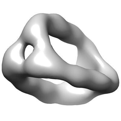 emd_8365.png emd_8365.png | 32.5 KB | ||
| Archive directory |  http://ftp.pdbj.org/pub/emdb/structures/EMD-8365ftp://ftp.pdbj.org/pub/emdb/structures/EMD-8365 http://ftp.pdbj.org/pub/emdb/structures/EMD-8365ftp://ftp.pdbj.org/pub/emdb/structures/EMD-8365 | HTTPS FTP |
-Related structure data


-Links
| EMDB pages | EMDB (EBI/PDBe) / EMDataResource |
|---|---|
| Related items in Molecule of the Month |

-Map
| File | Download / File: emd_8365.map.gz / Format: CCP4 / Size: 8 MB / Type: IMAGE STORED AS FLOATING POINT NUMBER (4 BYTES) | ||||||||||||||||||||||||||||||||||||||||||||||||||||||||||||||||||||
|---|---|---|---|---|---|---|---|---|---|---|---|---|---|---|---|---|---|---|---|---|---|---|---|---|---|---|---|---|---|---|---|---|---|---|---|---|---|---|---|---|---|---|---|---|---|---|---|---|---|---|---|---|---|---|---|---|---|---|---|---|---|---|---|---|---|---|---|---|---|
| Annotation | 10 nm RNA nanoprism | ||||||||||||||||||||||||||||||||||||||||||||||||||||||||||||||||||||
| Voxel size | X=Y=Z: 2.51 Å | ||||||||||||||||||||||||||||||||||||||||||||||||||||||||||||||||||||
| Density |
| ||||||||||||||||||||||||||||||||||||||||||||||||||||||||||||||||||||
| Symmetry | Space group: 1 | ||||||||||||||||||||||||||||||||||||||||||||||||||||||||||||||||||||
| Details | EMDB XML:
CCP4 map header:
| ||||||||||||||||||||||||||||||||||||||||||||||||||||||||||||||||||||
-Supplemental data
- Sample components
-Entire : 10 nm RNA prism
| Entire | Name: 10 nm RNA prism |
|---|---|
| Components |
|
-Supramolecule #1: 10 nm RNA prism
| Supramolecule | Name: 10 nm RNA prism / type: complex / ID: 1 / Parent: 0 / Macromolecule list: #1 Details: The RNA strands are made by in vitro transcription. |
|---|---|
| Source (natural) | Organism: synthetic construct (others) |
| Molecular weight | Experimental: 180 KDa |
-Experimental details
-Structure determination
| Method | cryo EM |
|---|---|
 Processing Processing | single particle reconstruction |
| Aggregation state | particle |
-Sample preparation
| Concentration | 0.2 mg/mL |
|---|---|
| Buffer | pH: 8 / Details: 100 mM NaCl, 50 mM Tris , 10 mM MgCl2 |
| Grid | Model: Quantifoil R1.2/1.3 / Material: COPPER / Mesh: 200 / Pretreatment - Type: GLOW DISCHARGE / Pretreatment - Atmosphere: AIR |
| Vitrification | Cryogen name: ETHANE / Chamber humidity: 100 % / Chamber temperature: 298 K / Instrument: FEI VITROBOT MARK IV Details: Blot for 3 seconds before plunging into liquid ethane (FEI VITROBOT MARK IV).. |
| Details | The RNA strands are made by in vitro transcription. |
- Electron microscopy
| Microscope | JEOL 2200FS |
|---|---|
| Electron beam | Acceleration voltage: 200 kV / Electron source: FIELD EMISSION GUN |
| Electron optics | Illumination mode: FLOOD BEAM / Imaging mode: BRIGHT FIELDBright-field microscopy / Cs: 2.0 mm / Nominal magnification: 25000 |
| Sample stage | Specimen holder model: GATAN 626 SINGLE TILT LIQUID NITROGEN CRYO TRANSFER HOLDER |
| Image recording | Film or detector model: DIRECT ELECTRON DE-20 (5k x 3k) / Detector mode: OTHER / Number real images: 52 / Average exposure time: 3.0 sec. / Average electron dose: 60.0 e/Å2 |
-Image processing
| Particle selection | Number selected: 2340 |
|---|---|
| CTF correction | Software - Name: EMAN (ver. 2.1) |
| Initial angle assignment | Type: NOT APPLICABLE |
| Final angle assignment | Type: COMMON LINE |
| Final reconstruction | Resolution.type: BY AUTHOR / Resolution: 25.0 Å / Resolution method: FSC 0.143 CUT-OFF / Software - Name: EMAN (ver. 2.1) / Number images used: 1514 |
