National Institutes of Health/National Institute Of Allergy and Infectious Diseases
AI79031
United States
National Institutes of Health/National Institute Of Allergy and Infectious Diseases
AI106005
United States
National Institutes of Health/National Institute Of Allergy and Infectious Diseases
AI123365
United States
Citation
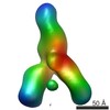
Journal: Proc Natl Acad Sci U S A / Year: 2016 Title: Structural flexibility at a major conserved antibody target on hepatitis C virus E2 antigen. Authors: Leopold Kong / David E Lee / Rameshwar U Kadam / Tong Liu / Erick Giang / Travis Nieusma / Fernando Garces / Netanel Tzarum / Virgil L Woods / Andrew B Ward / Sheng Li / Ian A Wilson / Mansun Law / Abstract: Hepatitis C virus (HCV) is a major cause of liver disease, affecting over 2% of the world's population. The HCV envelope glycoproteins E1 and E2 mediate viral entry, with E2 being the main target of ...Hepatitis C virus (HCV) is a major cause of liver disease, affecting over 2% of the world's population. The HCV envelope glycoproteins E1 and E2 mediate viral entry, with E2 being the main target of neutralizing antibody responses. Structural investigations of E2 have produced templates for vaccine design, including the conserved CD81 receptor-binding site (CD81bs) that is a key target of broadly neutralizing antibodies (bNAbs). Unfortunately, immunization with recombinant E2 and E1E2 rarely elicits sufficient levels of bNAbs for protection. To understand the challenges for eliciting bNAb responses against the CD81bs, we investigated the E2 CD81bs by electron microscopy (EM), hydrogen-deuterium exchange (HDX), molecular dynamics (MD), and calorimetry. By EM, we observed that HCV1, a bNAb recognizing the N-terminal region of the CD81bs, bound a soluble E2 core construct from multiple angles of approach, suggesting components of the CD81bs are flexible. HDX of multiple E2 constructs consistently indicated the entire CD81bs was flexible relative to the rest of the E2 protein, which was further confirmed by MD simulations. However, E2 has a high melting temperature of 84.8 °C, which is more akin to proteins from thermophilic organisms. Thus, recombinant E2 is a highly stable protein overall, but with an exceptionally flexible CD81bs. Such flexibility may promote induction of nonneutralizing antibodies over bNAbs to E2 CD81bs, underscoring the necessity of rigidifying this antigenic region as a target for rational vaccine design.
History
Deposition
Aug 16, 2016
-
Header (metadata) release
Aug 31, 2016
-
Map release
Oct 26, 2016
-
Update
Sep 6, 2023
-
Current status
Sep 6, 2023
Processing site: RCSB / Status: Released
-
Structure visualization
Movie
Surface view with section colored by density value
In the structure databanks used in Yorodumi, some data are registered as the other names, "COVID-19 virus" and "2019-nCoV". Here are the details of the virus and the list of structure data.
Jan 31, 2019. EMDB accession codes are about to change! (news from PDBe EMDB page)
EMDB accession codes are about to change! (news from PDBe EMDB page)
The allocation of 4 digits for EMDB accession codes will soon come to an end. Whilst these codes will remain in use, new EMDB accession codes will include an additional digit and will expand incrementally as the available range of codes is exhausted. The current 4-digit format prefixed with “EMD-” (i.e. EMD-XXXX) will advance to a 5-digit format (i.e. EMD-XXXXX), and so on. It is currently estimated that the 4-digit codes will be depleted around Spring 2019, at which point the 5-digit format will come into force.
The EM Navigator/Yorodumi systems omit the EMD- prefix.
Related info.:Q: What is EMD? / ID/Accession-code notation in Yorodumi/EM Navigator
Yorodumi is a browser for structure data from EMDB, PDB, SASBDB, etc.
This page is also the successor to EM Navigator detail page, and also detail information page/front-end page for Omokage search.
The word "yorodu" (or yorozu) is an old Japanese word meaning "ten thousand". "mi" (miru) is to see.
Related info.:EMDB / PDB / SASBDB / Comparison of 3 databanks / Yorodumi Search / Aug 31, 2016. New EM Navigator & Yorodumi / Yorodumi Papers / Jmol/JSmol / Function and homology information / Changes in new EM Navigator and Yorodumi
 Movie
Movie Controller


 Open data
 Basic information
 Map data
 Sample
 Keywords
 neutralizing antibodies /
  Hepatitis C virus /
 Authors
 United States, 3 items
  Citation
 Structure visualization
 Movie viewer
 Downloads & links
 emd_8339.png
 http://ftp.pdbj.org/pub/emdb/structures/EMD-8339
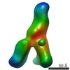


 Sample components
 Processing
 Electron microscopy
