 Movie
Movie Controller

[English] 日本語
 Yorodumi
- EMDB-8331: Glycan shield and epitope masking of a coronavirus spike protein ... -
+ Open data

- Basic information
| Entry | Database: EMDB / ID: EMD-8331 | |||||||||
|---|---|---|---|---|---|---|---|---|---|---|
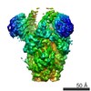
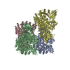
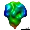
| Title | Glycan shield and epitope masking of a coronavirus spike protein observed by cryo-electron microscopy | |||||||||
 Map data Map data | Coronavirus spike protein | |||||||||
 Sample Sample |
| |||||||||
| Function / homology |  Function and homology information Function and homology informationendocytosis involved in viral entry into host cell / host cell endoplasmic reticulum-Golgi intermediate compartment membrane / receptor-mediated virion attachment to host cell / host cell surface receptor binding / fusion of virus membrane with host plasma membrane / fusion of virus membrane with host endosome membrane / viral envelope / virion membrane / membraneSimilarity search - Function | |||||||||
| Biological species |  Human coronavirus NL63 Human coronavirus NL63 | |||||||||
| Method | single particle reconstruction / cryo EM / Resolution: 3.4 Å | |||||||||
 Authors Authors | Walls AC / Tortorici MA / Frenz B / Snijder J / Li W / Rey FA / DiMaio F / Bosch BJ / Veesler D | |||||||||
| Funding support |  United States, 1 items United States, 1 items
| |||||||||
 Citation Citation | Journal: Nat Struct Mol Biol / Year: 2016 Title: Glycan shield and epitope masking of a coronavirus spike protein observed by cryo-electron microscopy. Authors: Alexandra C Walls / M Alejandra Tortorici / Brandon Frenz / Joost Snijder / Wentao Li / Félix A Rey / Frank DiMaio / Berend-Jan Bosch / David Veesler /   Abstract: The threat of a major coronavirus pandemic urges the development of strategies to combat these pathogens. Human coronavirus NL63 (HCoV-NL63) is an α-coronavirus that can cause severe lower- ...The threat of a major coronavirus pandemic urges the development of strategies to combat these pathogens. Human coronavirus NL63 (HCoV-NL63) is an α-coronavirus that can cause severe lower-respiratory-tract infections requiring hospitalization. We report here the 3.4-Å-resolution cryo-EM reconstruction of the HCoV-NL63 coronavirus spike glycoprotein trimer, which mediates entry into host cells and is the main target of neutralizing antibodies during infection. The map resolves the extensive glycan shield obstructing the protein surface and, in combination with mass spectrometry, provides a structural framework to understand the accessibility to antibodies. The structure reveals the complete architecture of the fusion machinery including the triggering loop and the C-terminal domains, which contribute to anchoring the trimer to the viral membrane. Our data further suggest that HCoV-NL63 and other coronaviruses use molecular trickery, based on epitope masking with glycans and activating conformational changes, to evade the immune system of infected hosts. | |||||||||
| History |
|
- Structure visualization
| Movie |
Movie viewer |
|---|---|
| Structure viewer | EM map: SurfViewMolmilJmol/JSmol |
| Supplemental images |

- Downloads & links
-EMDB archive
| Map data | emd_8331.map.gz | 10.7 MB | EMDB map data format | |
|---|---|---|---|---|
| Header (meta data) | emd-8331-v30.xmlemd-8331.xml | 14.6 KB 14.6 KB | Display Display | EMDB header |
| Images | 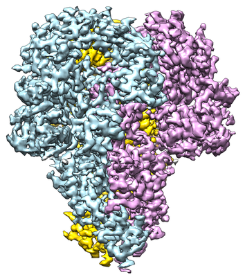 emd_8331.png emd_8331.png | 230.5 KB | ||
| Archive directory |  http://ftp.pdbj.org/pub/emdb/structures/EMD-8331ftp://ftp.pdbj.org/pub/emdb/structures/EMD-8331 http://ftp.pdbj.org/pub/emdb/structures/EMD-8331ftp://ftp.pdbj.org/pub/emdb/structures/EMD-8331 | HTTPS FTP |
-Related structure data
| Related structure data |  5szsMC M: atomic model generated by this map C: citing same article ( |
|---|---|
| Similar structure data |


-Links
| EMDB pages | EMDB (EBI/PDBe) / EMDataResource |
|---|
-Map
| File | Download / File: emd_8331.map.gz / Format: CCP4 / Size: 125 MB / Type: IMAGE STORED AS FLOATING POINT NUMBER (4 BYTES) | ||||||||||||||||||||||||||||||||||||||||||||||||||||||||||||||||||||
|---|---|---|---|---|---|---|---|---|---|---|---|---|---|---|---|---|---|---|---|---|---|---|---|---|---|---|---|---|---|---|---|---|---|---|---|---|---|---|---|---|---|---|---|---|---|---|---|---|---|---|---|---|---|---|---|---|---|---|---|---|---|---|---|---|---|---|---|---|---|
| Annotation | Coronavirus spike protein | ||||||||||||||||||||||||||||||||||||||||||||||||||||||||||||||||||||
| Voxel size | X=Y=Z: 1.36 Å | ||||||||||||||||||||||||||||||||||||||||||||||||||||||||||||||||||||
| Density |
| ||||||||||||||||||||||||||||||||||||||||||||||||||||||||||||||||||||
| Symmetry | Space group: 1 | ||||||||||||||||||||||||||||||||||||||||||||||||||||||||||||||||||||
| Details | EMDB XML:
CCP4 map header:
| ||||||||||||||||||||||||||||||||||||||||||||||||||||||||||||||||||||
-Supplemental data
- Sample components
-Entire : Human coronavirus NL63 spike glycoprotein ectodomain
| Entire | Name: Human coronavirus NL63 spike glycoprotein ectodomain |
|---|---|
| Components |
|
-Supramolecule #1: Human coronavirus NL63 spike glycoprotein ectodomain
| Supramolecule | Name: Human coronavirus NL63 spike glycoprotein ectodomain / type: complex / ID: 1 / Parent: 0 / Macromolecule list: #1 |
|---|---|
| Source (natural) | Organism: Human coronavirus NL63 |
| Recombinant expression | Organism:  Drosophila melanogaster (fruit fly) / Recombinant cell: Schneider 2 / Recombinant plasmid: pMT-BiP-V5-His Drosophila melanogaster (fruit fly) / Recombinant cell: Schneider 2 / Recombinant plasmid: pMT-BiP-V5-His |
| Molecular weight | Theoretical: 450 KDa |
-Macromolecule #1: Spike glycoprotein
| Macromolecule | Name: Spike glycoprotein / type: protein_or_peptide / ID: 1 / Number of copies: 3 / Enantiomer: LEVO |
|---|---|
| Source (natural) | Organism: Human coronavirus NL63 |
| Molecular weight | Theoretical: 146.813984 KDa |
| Recombinant expression | Organism: Drosophila melanogaster (fruit fly) |
| Sequence | String: FFTCNSNANL SMLQLGVPDN SSTIVTGLLP THWFCANQST SVYSANGFFY IDVGNHRSAF ALHTGYYDAN QYYIYVTNEI GLNASVTLK ICKFSRNTTF DFLSNASSSF DCIVNLLFTE QLGAPLGITI SGETVRLHLY NVTRTFYVPA AYKLTKLSVK C YFNYSCVF ...String: FFTCNSNANL SMLQLGVPDN SSTIVTGLLP THWFCANQST SVYSANGFFY IDVGNHRSAF ALHTGYYDAN QYYIYVTNEI GLNASVTLK ICKFSRNTTF DFLSNASSSF DCIVNLLFTE QLGAPLGITI SGETVRLHLY NVTRTFYVPA AYKLTKLSVK C YFNYSCVF SVVNATVTVN VTTHNGRVVN YTVCDDCNGY TDNIFSVQQD GRIPNGFPFN NWFLLTNGST LVDGVSRLYQ PL RLTCLWP VPGLKSSTGF VYFNATGSDV NCNGYQHNSV VDVMRYNLNF SANSLDNLKS GVIVFKTLQY DVLFYCSNSS SGV LDTTIP FGPSSQPYYC FINSTINTTH VSTFVGILPP TVREIVVART GQFYINGFKY FDLGFIEAVN FNVTTASATD FWTV AFATF VDVLVNVSAT NIQNLLYCDS PFEKLQCEHL QFGLQDGFYS ANFLDDNVLP ETYVALPIYY QHTDINFTAT ASFGG SCYV CKPHQVNISL NGNTSVCVRT SHFSIRYIYN RVKSGSPGDS SWHIYLKSGT CPFSFSKLNN FQKFKTICFS TVEVPG SCN FPLEATWHYT SYTIVGALYV TWSEGNSITG VPYPVSGIRE FSNLVLNNCT KYNIYDYVGT GIIRSSNQSL AGGITYV SN SGNLLGFKNV STGNIFIVTP CNQPDQVAVY QQSIIGAMTA VNESRYGLQN LLQLPNFYYV SNGGNNCTTA VMTYSNFG I CADGSLIPVR PRNSSDNGIS AIITANLSIP SNWTTSVQVE YLQITSTPIV VDCATYVCNG NPRCKNLLKQ YTSACKTIE DALRLSAHLE TNDVSSMLTF DSNAFSLANV TSFGDYNLSS VLPQRNIRSS RIAGRSALED LLFSKVVTSG LGTVDVDYKS CTKGLSIAD LACAQYYNGI MVLPGVADAE RMAMYTGSLI GGMVLGGLTS AAAIPFSLAL QARLNYVALQ TDVLQENQKI L AASFNKAI NNIVASFSSV NDAITQTAEA IHTVTIALNK IQDVVNQQGS ALNHLTSQLR HNFQAISNSI QAIYDRLDSI QA DQQVDRL ITGRLAALNA FVSQVLNKYT EVRGSRRLAQ QKINECVKSQ SNRYGFCGNG THIFSIVNSA PDGLLFLHTV LLP TDYKNV KAWSGICVDG IYGYVLRQPN LVLYSDNGVF RVTSRVMFQP RLPVLSDFVQ IYNCNVTFVN ISRVELHTVI PDYV DVNKT LQEFAQNLPK YVKPNFDLTP FNLTYLNLSS ELKQLEAKTA SLFQTTVELQ GLIDQINSTY VDLKLLNRFE NLIKR MKQI EDKIEEIESK QKKIENEIAR IKKIKLVPRG SLEWSHPQFE K |
-Macromolecule #9: 2-acetamido-2-deoxy-beta-D-glucopyranose
| Macromolecule | Name: 2-acetamido-2-deoxy-beta-D-glucopyranose / type: ligand / ID: 9 / Number of copies: 21 / Formula: NAG |
|---|---|
| Molecular weight | Theoretical: 221.208 Da |
| Chemical component information |  ChemComp-NAG: |
-Experimental details
-Structure determination
| Method | cryo EM |
|---|---|
 Processing Processing | single particle reconstruction |
| Aggregation state | particle |
-Sample preparation
| Concentration | 1 mg/mL | ||||||
|---|---|---|---|---|---|---|---|
| Buffer | pH: 7.5 Component:
| ||||||
| Grid | Model: Protochips C-flat 1.2/1.3 / Material: COPPER / Mesh: 400 / Pretreatment - Type: GLOW DISCHARGE / Details: 20 mA | ||||||
| Vitrification | Cryogen name: ETHANE |
- Electron microscopy
| Microscope | FEI TITAN KRIOS |
|---|---|
| Electron beam | Acceleration voltage: 300 kV / Electron source: FIELD EMISSION GUN |
| Electron optics | Illumination mode: FLOOD BEAM / Imaging mode: BRIGHT FIELDBright-field microscopy |
| Image recording | Film or detector model: GATAN K2 SUMMIT (4k x 4k) / Detector mode: COUNTING / Digitization - Sampling interval: 5.0 µm / Digitization - Frames/image: 1-50 / Average exposure time: 10.0 sec. / Average electron dose: 48.0 e/Å2 |
| Experimental equipment |  Model: Titan Krios / Image courtesy: FEI Company |
-Image processing
| Particle selection | Number selected: 474000 |
|---|---|
| CTF correction | Software - Name: GCTF/Relion |
| Startup model | Type of model: OTHER Details: Common-lines generated model for the MHV spike glycoprotein |
| Initial angle assignment | Type: PROJECTION MATCHING / Software - Name: RELION |
| Final 3D classification | Software - Name: RELION |
| Final angle assignment | Type: PROJECTION MATCHING / Software - Name: RELION |
| Final reconstruction | Applied symmetry - Point group: C3 (3 fold cyclic) / Algorithm: FOURIER SPACE / Resolution.type: BY AUTHOR / Resolution: 3.4 Å / Resolution method: FSC 0.143 CUT-OFF / Software - Name: RELION / Number images used: 79667 |
-Atomic model buiding 1
| Refinement | Space: REAL / Protocol: AB INITIO MODEL |
|---|---|
| Output model | PDB-5szs: |
