National Institutes of Health/National Institute of Biomedical Imaging and Bioengineering (NIH/NIBIB)
EB001567
United States
Citation
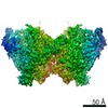
Journal: Proc Natl Acad Sci U S A / Year: 2016 Title: Archaeal flagellin combines a bacterial type IV pilin domain with an Ig-like domain. Authors: Tatjana Braun / Matthijn R Vos / Nir Kalisman / Nicholas E Sherman / Reinhard Rachel / Reinhard Wirth / Gunnar F Schröder / Edward H Egelman / Abstract: The bacterial flagellar apparatus, which involves ∼40 different proteins, has been a model system for understanding motility and chemotaxis. The bacterial flagellar filament, largely composed of a ...The bacterial flagellar apparatus, which involves ∼40 different proteins, has been a model system for understanding motility and chemotaxis. The bacterial flagellar filament, largely composed of a single protein, flagellin, has been a model for understanding protein assembly. This system has no homology to the eukaryotic flagellum, in which the filament alone, composed of a microtubule-based axoneme, contains more than 400 different proteins. The archaeal flagellar system is simpler still, in some cases having ∼13 different proteins with a single flagellar filament protein. The archaeal flagellar system has no homology to the bacterial one and must have arisen by convergent evolution. However, it has been understood that the N-terminal domain of the archaeal flagellin is a homolog of the N-terminal domain of bacterial type IV pilin, showing once again how proteins can be repurposed in evolution for different functions. Using cryo-EM, we have been able to generate a nearly complete atomic model for a flagellar-like filament of the archaeon Ignicoccus hospitalis from a reconstruction at ∼4-Å resolution. We can now show that the archaeal flagellar filament contains a β-sandwich, previously seen in the FlaF protein that forms the anchor for the archaeal flagellar filament. In contrast to the bacterial flagellar filament, where the outer globular domains make no contact with each other and are not necessary for either assembly or motility, the archaeal flagellin outer domains make extensive contacts with each other that largely determine the interesting mechanical properties of these filaments, allowing these filaments to flex.
History
Deposition
Aug 23, 2016
-
Header (metadata) release
Sep 7, 2016
-
Map release
Sep 7, 2016
-
Update
Dec 11, 2019
-
Current status
Dec 11, 2019
Processing site: RCSB / Status: Released
-
Structure visualization
Movie
Surface view with section colored by density value
In the structure databanks used in Yorodumi, some data are registered as the other names, "COVID-19 virus" and "2019-nCoV". Here are the details of the virus and the list of structure data.
Jan 31, 2019. EMDB accession codes are about to change! (news from PDBe EMDB page)
EMDB accession codes are about to change! (news from PDBe EMDB page)
The allocation of 4 digits for EMDB accession codes will soon come to an end. Whilst these codes will remain in use, new EMDB accession codes will include an additional digit and will expand incrementally as the available range of codes is exhausted. The current 4-digit format prefixed with “EMD-” (i.e. EMD-XXXX) will advance to a 5-digit format (i.e. EMD-XXXXX), and so on. It is currently estimated that the 4-digit codes will be depleted around Spring 2019, at which point the 5-digit format will come into force.
The EM Navigator/Yorodumi systems omit the EMD- prefix.
Related info.:Q: What is EMD? / ID/Accession-code notation in Yorodumi/EM Navigator
Yorodumi is a browser for structure data from EMDB, PDB, SASBDB, etc.
This page is also the successor to EM Navigator detail page, and also detail information page/front-end page for Omokage search.
The word "yorodu" (or yorozu) is an old Japanese word meaning "ten thousand". "mi" (miru) is to see.
Related info.:EMDB / PDB / SASBDB / Comparison of 3 databanks / Yorodumi Search / Aug 31, 2016. New EM Navigator & Yorodumi / Yorodumi Papers / Jmol/JSmol / Function and homology information / Changes in new EM Navigator and Yorodumi
 Movie
Movie Controller


 Open data
 Basic information
 Map data
 Sample
 membrane / Archaeal Type IV pilin N-terminal domain-containing protein
 Function and homology information

 Authors
 United States, 1 items
  Citation


 Structure visualization
 Downloads & links
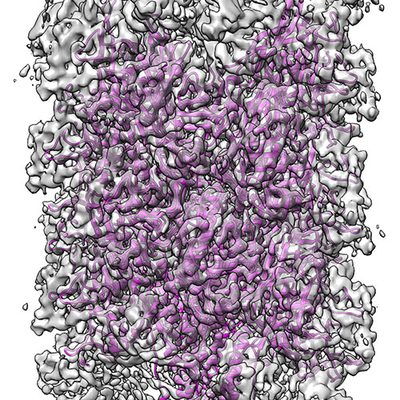 emd_8298.png
 http://ftp.pdbj.org/pub/emdb/structures/EMD-8298


 Sample components
 Processing
 Electron microscopy
