 Movie
Movie Controller


+ Open data

- Basic information
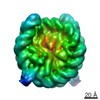
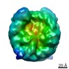
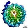
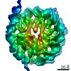
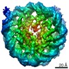
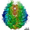
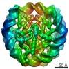
| Entry | Database: EMDB / ID: EMD-8247 | |||||||||
|---|---|---|---|---|---|---|---|---|---|---|
| Title | 53BP1 bound to a ubiquitylated and methylated nucleosome | |||||||||
 Map data Map data | NCP-ubme | |||||||||
 Sample Sample |
| |||||||||
| Biological species | synthetic construct (others) /  Xenopus laevis (African clawed frog) / Xenopus laevis (African clawed frog) /  Homo sapiens (human) Homo sapiens (human) | |||||||||
| Method | single particle reconstruction / cryo EM / Resolution: 7.73 Å | |||||||||
 Authors Authors | Wilson MD / Benlekbir S / Sicheri F / Rubinstein JL / Durocher D | |||||||||
 Citation Citation | Journal: Nature / Year: 2016 Title: The structural basis of modified nucleosome recognition by 53BP1. Abstract: DNA double-strand breaks (DSBs) elicit a histone modification cascade that controls DNA repair. This pathway involves the sequential ubiquitination of histones H1 and H2A by the E3 ubiquitin ligases ...DNA double-strand breaks (DSBs) elicit a histone modification cascade that controls DNA repair. This pathway involves the sequential ubiquitination of histones H1 and H2A by the E3 ubiquitin ligases RNF8 and RNF168, respectively. RNF168 ubiquitinates H2A on lysine 13 and lysine 15 (refs 7, 8) (yielding H2AK13ub and H2AK15ub, respectively), an event that triggers the recruitment of 53BP1 (also known as TP53BP1) to chromatin flanking DSBs. 53BP1 binds specifically to H2AK15ub-containing nucleosomes through a peptide segment termed the ubiquitination-dependent recruitment motif (UDR), which requires the simultaneous engagement of histone H4 lysine 20 dimethylation (H4K20me2) by its tandem Tudor domain. How 53BP1 interacts with these two histone marks in the nucleosomal context, how it recognizes ubiquitin, and how it discriminates between H2AK13ub and H2AK15ub is unknown. Here we present the electron cryomicroscopy (cryo-EM) structure of a dimerized human 53BP1 fragment bound to a H4K20me2-containing and H2AK15ub-containing nucleosome core particle (NCP-ubme) at 4.5 Å resolution. The structure reveals that H4K20me2 and H2AK15ub recognition involves intimate contacts with multiple nucleosomal elements including the acidic patch. Ubiquitin recognition by 53BP1 is unusual and involves the sandwiching of the UDR segment between ubiquitin and the NCP surface. The selectivity for H2AK15ub is imparted by two arginine fingers in the H2A amino-terminal tail, which straddle the nucleosomal DNA and serve to position ubiquitin over the NCP-bound UDR segment. The structure of the complex between NCP-ubme and 53BP1 reveals the basis of 53BP1 recruitment to DSB sites and illuminates how combinations of histone marks and nucleosomal elements cooperate to produce highly specific chromatin responses, such as those elicited following chromosome breaks. | |||||||||
| History |
|
- Structure visualization
| Movie |
 Movie viewer Movie viewer |
|---|---|
| Structure viewer | EM map: SurfViewMolmilJmol/JSmol |
| Supplemental images |

- Downloads & links
-EMDB archive
| Map data | emd_8247.map.gz | 7.2 MB | EMDB map data format | |
|---|---|---|---|---|
| Header (meta data) | emd-8247-v30.xmlemd-8247.xml | 17.4 KB 17.4 KB | Display Display | EMDB header |
| FSC (resolution estimation) | emd_8247_fsc.xml | 4.7 KB | Display | FSC data file |
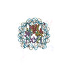
| Images | 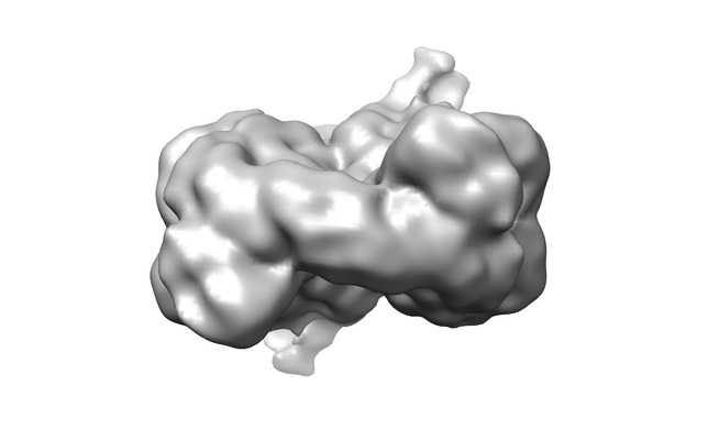 emd_8247.png emd_8247.png | 54.9 KB | ||
| Archive directory |  http://ftp.pdbj.org/pub/emdb/structures/EMD-8247ftp://ftp.pdbj.org/pub/emdb/structures/EMD-8247 http://ftp.pdbj.org/pub/emdb/structures/EMD-8247ftp://ftp.pdbj.org/pub/emdb/structures/EMD-8247 | HTTPS FTP |
-Related structure data

-Links
| EMDB pages | EMDB (EBI/PDBe) / EMDataResource |
|---|
-Map
| File | Download / File: emd_8247.map.gz / Format: CCP4 / Size: 8 MB / Type: IMAGE STORED AS FLOATING POINT NUMBER (4 BYTES) | ||||||||||||||||||||||||||||||||||||||||||||||||||||||||||||||||||||
|---|---|---|---|---|---|---|---|---|---|---|---|---|---|---|---|---|---|---|---|---|---|---|---|---|---|---|---|---|---|---|---|---|---|---|---|---|---|---|---|---|---|---|---|---|---|---|---|---|---|---|---|---|---|---|---|---|---|---|---|---|---|---|---|---|---|---|---|---|---|
| Annotation | NCP-ubme | ||||||||||||||||||||||||||||||||||||||||||||||||||||||||||||||||||||
| Voxel size | X=Y=Z: 1.45 Å | ||||||||||||||||||||||||||||||||||||||||||||||||||||||||||||||||||||
| Density |
| ||||||||||||||||||||||||||||||||||||||||||||||||||||||||||||||||||||
| Symmetry | Space group: 1 | ||||||||||||||||||||||||||||||||||||||||||||||||||||||||||||||||||||
| Details | EMDB XML:
CCP4 map header:
| ||||||||||||||||||||||||||||||||||||||||||||||||||||||||||||||||||||
-Supplemental data
- Sample components
-Entire : NCP-ubme
| Entire | Name: NCP-ubme |
|---|---|
| Components |
|
-Supramolecule #1: NCP-ubme
| Supramolecule | Name: NCP-ubme / type: complex / ID: 1 / Parent: 0 Details: Recombinant ubiquitylated and methylated nucleosome core particle |
|---|---|
| Molecular weight | Theoretical: 218 KDa |
-Supramolecule #2: NCP-ubme
| Supramolecule | Name: NCP-ubme / type: complex / ID: 2 / Parent: 1 Details: Modified nuclesome core particles, H2A enzymatically ubiquitylated on H2A K15, H4 chemically alkylated at K20C to create dimethyl lysine analog |
|---|---|
| Molecular weight | Theoretical: 226 KDa |
-Supramolecule #3: Widom-601 DNA
| Supramolecule | Name: Widom-601 DNA / type: complex / ID: 3 / Parent: 2 Details: 145 bp fragment of Widom-601 strong nucleosome positioning DNA sequence, gift from Curt Davey (Vasudevan et. al, 2010, J.Mol.Biol.) |
|---|---|
| Source (natural) | Organism: synthetic construct (others) |
| Recombinant expression | Organism:  Escherichia coli (E. coli) / Recombinant plasmid: pUC57-8x145bp Escherichia coli (E. coli) / Recombinant plasmid: pUC57-8x145bp |
| Molecular weight | Theoretical: 882.31 KDa |
-Supramolecule #4: H4Kc20me2
| Supramolecule | Name: H4Kc20me2 / type: complex / ID: 4 / Parent: 2 Details: Histone H4 alkylated at position 20 to create dimethyl lysine analog |
|---|---|
| Source (natural) | Organism: Xenopus laevis (African clawed frog) |
| Recombinant expression | Organism: Escherichia coli (E. coli) / Recombinant plasmid: pDD2349 |
-Supramolecule #5: Histone H3
| Supramolecule | Name: Histone H3 / type: complex / ID: 5 / Parent: 2 |
|---|---|
| Source (natural) | Organism: Xenopus laevis (African clawed frog) |
| Recombinant expression | Organism: Escherichia coli (E. coli) / Recombinant plasmid: pDD1874 |
-Supramolecule #6: Histone H2A.1 K13RK36RT16S
| Supramolecule | Name: Histone H2A.1 K13RK36RT16S / type: complex / ID: 6 / Parent: 2 Details: Histone H2A.1 covalently cross-linked at K15 to ubiquitin at G76 by an isopeptide bond |
|---|---|
| Source (natural) | Organism: Homo sapiens (human) |
| Recombinant expression | Organism: Escherichia coli (E. coli) / Recombinant plasmid: pDD2647 |
-Supramolecule #7: Histone H2B.1
| Supramolecule | Name: Histone H2B.1 / type: complex / ID: 7 / Parent: 2 |
|---|---|
| Source (natural) | Organism: Homo sapiens (human) |
| Recombinant expression | Organism: Escherichia coli (E. coli) / Recombinant plasmid: pDD2644 |
-Experimental details
-Structure determination
| Method | cryo EM |
|---|---|
 Processing Processing | single particle reconstruction |
| Aggregation state | particle |
-Sample preparation
| Concentration | 0.6 mg/mL | |||||||||||||||
|---|---|---|---|---|---|---|---|---|---|---|---|---|---|---|---|---|
| Buffer | pH: 7.5 Component:
Details: High concentration NCP-ubme/GST-53BP1 complex in 200 mM salt was diluted just prior to grid freezing. | |||||||||||||||
| Grid | Model: electron micrsocopy sciences / Material: COPPER/RHODIUM / Mesh: 400 / Support film - Material: CARBON / Support film - topology: HOLEY / Support film - Film thickness: 40.0 nm / Pretreatment - Type: GLOW DISCHARGE / Pretreatment - Atmosphere: AIR / Pretreatment - Pressure: 0.039 kPa | |||||||||||||||
| Vitrification | Cryogen name: ETHANE-PROPANE / Chamber humidity: 100 % / Chamber temperature: 277 K / Instrument: FEI VITROBOT MARK III Details: Plunged into liquid ethane-propane (FEI VITROBOT MARK III). |
- Electron microscopy
| Microscope | FEI TECNAI F20 |
|---|---|
| Electron beam | Acceleration voltage: 200 kV / Electron source: FIELD EMISSION GUN |
| Electron optics | C2 aperture diameter: 30.0 µm / Calibrated magnification: 34483 / Illumination mode: FLOOD BEAM / Imaging mode: BRIGHT FIELDBright-field microscopy / Cs: 2.0 mm / Nominal magnification: 25000 |
| Sample stage | Specimen holder model: GATAN 626 SINGLE TILT LIQUID NITROGEN CRYO TRANSFER HOLDER Cooling holder cryogen: NITROGEN |
| Image recording | Film or detector model: GATAN K2 SUMMIT (4k x 4k) / Detector mode: COUNTING / Digitization - Frames/image: 1-30 / Number grids imaged: 2 / Number real images: 227 / Average exposure time: 0.5 sec. / Average electron dose: 36.0 e/Å2 |
| Experimental equipment |  Model: Tecnai F20 / Image courtesy: FEI Company |
-Image processing
| Particle selection | Number selected: 52843 Details: Automatically picked from roughly 1000 particles using a manually picked template |
|---|---|
| CTF correction | Software - Name: CTFFIND (ver. 3) |
| Startup model | Type of model: PDB ENTRY PDB model - PDB ID: Details: Resized to the same pixel size as the data and high-pass filtered to 40 Angstrom resolution |
| Initial angle assignment | Type: ANGULAR RECONSTITUTION / Software - Name: RELION (ver. 1.3) |
| Final 3D classification | Number classes: 4 / Software - Name: RELION (ver. 1.3) |
| Final angle assignment | Type: ANGULAR RECONSTITUTION / Software - Name: RELION (ver. 1.3) |
| Final reconstruction | Number classes used: 2 / Applied symmetry - Point group: C2 (2 fold cyclic) / Algorithm: FOURIER SPACE / Resolution.type: BY AUTHOR / Resolution: 7.73 Å / Resolution method: FSC 0.143 CUT-OFF / Software - Name: RELION (ver. 1.3) / Number images used: 10133 |
| FSC plot (resolution estimation) |  |
-Atomic model buiding 1
| Details | Local fitting was performed in Chimera using rigid body fitting. No associate co-ordinates were deposited. |
|---|---|
| Refinement | Space: REAL / Protocol: RIGID BODY FIT / Overall B value: 1 |
