 Movie
Movie Controller

[English] 日本語
 Yorodumi
- EMDB-8199: Structure of GNNQQNY from yeast prion Sup35 in space group P21212... -
+ Open data

- Basic information
| Entry | Database: EMDB / ID: EMD-8199 | |||||||||||||||||||||
|---|---|---|---|---|---|---|---|---|---|---|---|---|---|---|---|---|---|---|---|---|---|---|
| Title | Structure of GNNQQNY from yeast prion Sup35 in space group P212121 determined by MicroED | |||||||||||||||||||||
 Map data Map data | GNNQQNY from yeast prion Sup35 | |||||||||||||||||||||
 Sample Sample |
| |||||||||||||||||||||
 Keywords Keywords |  amyloid / yeast prion / PROTEIN FIBRIL amyloid / yeast prion / PROTEIN FIBRIL | |||||||||||||||||||||
| Function / homology |  Function and homology information Function and homology informationEukaryotic Translation Termination / translation release factor complex / translation release factor activity / nuclear-transcribed mRNA catabolic process, deadenylation-dependent decay / Nonsense Mediated Decay (NMD) independent of the Exon Junction Complex (EJC) / Nonsense Mediated Decay (NMD) enhanced by the Exon Junction Complex (EJC) / translational termination / Hydrolases; Acting on acid anhydrides; Acting on GTP to facilitate cellular and subcellular movement / cytoplasmic stress granule / translation ...Eukaryotic Translation Termination / translation release factor complex / translation release factor activity / nuclear-transcribed mRNA catabolic process, deadenylation-dependent decay / Nonsense Mediated Decay (NMD) independent of the Exon Junction Complex (EJC) / Nonsense Mediated Decay (NMD) enhanced by the Exon Junction Complex (EJC) / translational termination / Hydrolases; Acting on acid anhydrides; Acting on GTP to facilitate cellular and subcellular movement / cytoplasmic stress granule / translation / mRNA binding / GTPase activity / GTP binding / identical protein binding / cytosol / cytoplasmSimilarity search - Function | |||||||||||||||||||||
| Biological species |  Saccharomyces cerevisiae (brewer's yeast) Saccharomyces cerevisiae (brewer's yeast) | |||||||||||||||||||||
| Method | electron crystallography / cryo EM | |||||||||||||||||||||
 Authors Authors | Rodriguez JA / Sawaya MR | |||||||||||||||||||||
| Funding support |  United States, 6 items United States, 6 items
| |||||||||||||||||||||
 Citation Citation | Journal: Proc Natl Acad Sci U S A / Year: 2016 Title: Ab initio structure determination from prion nanocrystals at atomic resolution by MicroED. Authors: Michael R Sawaya / Jose Rodriguez / Duilio Cascio / Michael J Collazo / Dan Shi / Francis E Reyes / Johan Hattne / Tamir Gonen / David S Eisenberg / Abstract: Electrons, because of their strong interaction with matter, produce high-resolution diffraction patterns from tiny 3D crystals only a few hundred nanometers thick in a frozen-hydrated state. This ...Electrons, because of their strong interaction with matter, produce high-resolution diffraction patterns from tiny 3D crystals only a few hundred nanometers thick in a frozen-hydrated state. This discovery offers the prospect of facile structure determination of complex biological macromolecules, which cannot be coaxed to form crystals large enough for conventional crystallography or cannot easily be produced in sufficient quantities. Two potential obstacles stand in the way. The first is a phenomenon known as dynamical scattering, in which multiple scattering events scramble the recorded electron diffraction intensities so that they are no longer informative of the crystallized molecule. The second obstacle is the lack of a proven means of de novo phase determination, as is required if the molecule crystallized is insufficiently similar to one that has been previously determined. We show with four structures of the amyloid core of the Sup35 prion protein that, if the diffraction resolution is high enough, sufficiently accurate phases can be obtained by direct methods with the cryo-EM method microelectron diffraction (MicroED), just as in X-ray diffraction. The success of these four experiments dispels the concern that dynamical scattering is an obstacle to ab initio phasing by MicroED and suggests that structures of novel macromolecules can also be determined by direct methods. | |||||||||||||||||||||
| History |
|
- Structure visualization
| Movie |
Movie viewer |
|---|---|
| Structure viewer | EM map: SurfViewMolmilJmol/JSmol |
| Supplemental images |

- Downloads & links
-EMDB archive
| Map data | emd_8199.map.gz | 844.9 KB | EMDB map data format | |
|---|---|---|---|---|
| Header (meta data) | emd-8199-v30.xmlemd-8199.xml | 18.4 KB 18.4 KB | Display Display | EMDB header |
| Images | 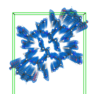 emd_8199.png emd_8199.png | 186.5 KB | ||
| Filedesc metadata | emd-8199.cif.gz | 5.7 KB | ||
| Filedesc structureFactors | emd_8199_sf.cif.gz | 39.8 KB | ||
| Archive directory |  http://ftp.pdbj.org/pub/emdb/structures/EMD-8199ftp://ftp.pdbj.org/pub/emdb/structures/EMD-8199 http://ftp.pdbj.org/pub/emdb/structures/EMD-8199ftp://ftp.pdbj.org/pub/emdb/structures/EMD-8199 | HTTPS FTP |
-Related structure data
| Related structure data |  5k2hMC  8196C  8197C 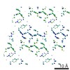 8198C  5k2eC  5k2fC  5k2gC C: citing same article ( M: atomic model generated by this map |
|---|---|
| Similar structure data |
-Links
| EMDB pages | EMDB (EBI/PDBe) / EMDataResource |
|---|---|
| Related items in Molecule of the Month |


-Map
| File | Download / File: emd_8199.map.gz / Format: CCP4 / Size: 2.2 MB / Type: IMAGE STORED AS FLOATING POINT NUMBER (4 BYTES) | ||||||||||||||||||||||||||||||||||||||||||||||||||||||||||||||||||||
|---|---|---|---|---|---|---|---|---|---|---|---|---|---|---|---|---|---|---|---|---|---|---|---|---|---|---|---|---|---|---|---|---|---|---|---|---|---|---|---|---|---|---|---|---|---|---|---|---|---|---|---|---|---|---|---|---|---|---|---|---|---|---|---|---|---|---|---|---|---|
| Annotation | GNNQQNY from yeast prion Sup35 | ||||||||||||||||||||||||||||||||||||||||||||||||||||||||||||||||||||
| Voxel size | X: 0.34059 Å / Y: 0.30813 Å / Z: 0.33758 Å | ||||||||||||||||||||||||||||||||||||||||||||||||||||||||||||||||||||
| Density |
| ||||||||||||||||||||||||||||||||||||||||||||||||||||||||||||||||||||
| Symmetry | Space group: 19 | ||||||||||||||||||||||||||||||||||||||||||||||||||||||||||||||||||||
| Details | EMDB XML:
CCP4 map header:
| ||||||||||||||||||||||||||||||||||||||||||||||||||||||||||||||||||||
-Supplemental data
- Sample components
-Entire : Prion fibril composed of a 7-residue segment of Sup35
| Entire | Name: Prion fibril composed of a 7-residue segment of Sup35 |
|---|---|
| Components |
|
-Supramolecule #1: Prion fibril composed of a 7-residue segment of Sup35
| Supramolecule | Name: Prion fibril composed of a 7-residue segment of Sup35 / type: complex / ID: 1 / Parent: 0 / Macromolecule list: #1 |
|---|---|
| Source (natural) | Organism: Saccharomyces cerevisiae (brewer's yeast) |
| Molecular weight | Theoretical: 3.25 kDa/nm |
-Macromolecule #1: Eukaryotic peptide chain release factor GTP-binding subunit
| Macromolecule | Name: Eukaryotic peptide chain release factor GTP-binding subunit type: protein_or_peptide / ID: 1 / Number of copies: 1 / Enantiomer: LEVO |
|---|---|
| Source (natural) | Organism: Saccharomyces cerevisiae (brewer's yeast) |
| Molecular weight | Theoretical: 836.807 Da |
| Sequence | String: GNNQQNY UniProtKB: Eukaryotic peptide chain release factor GTP-binding subunit |
-Macromolecule #2: water
| Macromolecule | Name: water / type: ligand / ID: 2 / Number of copies: 6 / Formula: HOH |
|---|---|
| Molecular weight | Theoretical: 18.015 Da |
| Chemical component information |  ChemComp-HOH: |
-Experimental details
-Structure determination
| Method | cryo EM |
|---|---|
 Processing Processing | electron crystallography |
| Aggregation state | 3D array |
-Sample preparation
| Concentration | 10 mg/mL |
|---|---|
| Buffer | pH: 7 / Details: water |
| Grid | Model: Quantifoil R2/2 / Material: COPPER / Mesh: 300 / Support film - Material: CARBON / Support film - topology: HOLEY ARRAY / Support film - Film thickness: 30 / Pretreatment - Type: GLOW DISCHARGE / Pretreatment - Time: 20 sec. / Pretreatment - Atmosphere: AIR |
| Vitrification | Cryogen name: ETHANE / Instrument: FEI VITROBOT MARK IV Details: Plunged into liquid ethane (FEI VITROBOT MARK IV). |
| Details | crystal |
| Crystal formation | Lipid mixture: none / Instrument: 24-well plate Atmosphere: in air, in sealed chamber, in equilibrium with reservoir solution Temperature: 298.0 K / Time: 1.0 DAY Details: Grown in batch at ~20 degrees C in a microcentrifuge tube. Crystals grew within a day after seeding with NNQQNY-Zn crystals. |
- Electron microscopy
| Microscope | FEI TECNAI F20 |
|---|---|
| Electron beam | Acceleration voltage: 200 kV / Electron source: FIELD EMISSION GUN |
| Electron optics | Illumination mode: FLOOD BEAM / Imaging mode: DIFFRACTION / Camera length: 1350 mm |
| Sample stage | Specimen holder model: GATAN 626 SINGLE TILT LIQUID NITROGEN CRYO TRANSFER HOLDER Cooling holder cryogen: NITROGEN |
| Temperature | Min: 100.0 K / Max: 100.0 K |
| Image recording | Film or detector model: TVIPS TEMCAM-F416 (4k x 4k) / Digitization - Dimensions - Width: 4096 pixel / Digitization - Dimensions - Height: 4096 pixel / Number grids imaged: 2 / Number diffraction images: 127 / Average exposure time: 2.0 sec. / Average electron dose: 0.01 e/Å2 Details: The detector was operated in rolling shutter mode with 2x2 pixel binning. |
| Experimental equipment |  Model: Tecnai F20 / Image courtesy: FEI Company |
-Image processing
| Crystallography statistics | Number intensities measured: 16753 / Number structure factors: 2399 / Fourier space coverage: 82.7 / R sym: 15.1 / R merge: 15.1 / Overall phase error: 0.1 / Overall phase residual: 0.1 / Phase error rejection criteria: 0 / High resolution: 1.0 Å Details: Phase statistics are not applicable. No imaging was used. The phases were obtained by a crystallographic direct methods program, SHELXD. Shell:
| ||||||||||||||||||||||||||||||||||||||||||
|---|---|---|---|---|---|---|---|---|---|---|---|---|---|---|---|---|---|---|---|---|---|---|---|---|---|---|---|---|---|---|---|---|---|---|---|---|---|---|---|---|---|---|---|
| Final reconstruction | Resolution method: DIFFRACTION PATTERN/LAYERLINES / Software - Name: SHELXD (ver. 2013/2) / Software - details: direct methods Details: The density map was obtained using measured diffraction intensities and phases acquired from crystallographic direct methods program SHELXD. | ||||||||||||||||||||||||||||||||||||||||||
| Merging software list | Software - Name: SCALEPACK (ver. 1.98.7) |
-Atomic model buiding 1
| Refinement | Space: RECIPROCAL / Protocol: OTHER / Overall B value: 4.7 / Target criteria: maximum likelihood |
|---|---|
| Output model | PDB-5k2h: |
