 Movie
Movie Controller

[English] 日本語
 Yorodumi
- EMDB-8084: The structure of microsomal glutathione transferase 1 in complex ... -
+ Open data

- Basic information
| Entry | Database: EMDB / ID: EMD-8084 | |||||||||
|---|---|---|---|---|---|---|---|---|---|---|
| Title | The structure of microsomal glutathione transferase 1 in complex with Meisenheimer complex | |||||||||
 Map data Map data | None | |||||||||
 Sample Sample |
| |||||||||
| Function / homology |  Function and homology information Function and homology informationcellular response to lipid hydroperoxide / Aflatoxin activation and detoxification / glutathione transport /  Glutathione conjugation / glutathione binding / Leydig cell differentiation / glutathione peroxidase activity / peroxisomal membrane / Neutrophil degranulation / glutathione transferase ...cellular response to lipid hydroperoxide / Aflatoxin activation and detoxification / glutathione transport / Glutathione conjugation / glutathione binding / Leydig cell differentiation / glutathione peroxidase activity / peroxisomal membrane / Neutrophil degranulation / glutathione transferase / glutathione transferase activity / glutathione metabolic process / response to organonitrogen compound / apical part of cell / mitochondrial outer membrane / response to lipopolysaccharide / response to xenobiotic stimulus / endoplasmic reticulum membrane / endoplasmic reticulum / mitochondrion / membrane / identical protein binding Glutathione conjugation / glutathione binding / Leydig cell differentiation / glutathione peroxidase activity / peroxisomal membrane / Neutrophil degranulation / glutathione transferase ...cellular response to lipid hydroperoxide / Aflatoxin activation and detoxification / glutathione transport / Glutathione conjugation / glutathione binding / Leydig cell differentiation / glutathione peroxidase activity / peroxisomal membrane / Neutrophil degranulation / glutathione transferase / glutathione transferase activity / glutathione metabolic process / response to organonitrogen compound / apical part of cell / mitochondrial outer membrane / response to lipopolysaccharide / response to xenobiotic stimulus / endoplasmic reticulum membrane / endoplasmic reticulum / mitochondrion / membrane / identical protein bindingSimilarity search - Function | |||||||||
| Biological species |  Rattus norvegicus (Norway rat) Rattus norvegicus (Norway rat) | |||||||||
| Method | electron crystallography / cryo EM / Resolution: 3.5 Å | |||||||||
 Authors Authors | Kuang Q / Purhonen P / Jegerschold C / Morgenstern R / Hebert H | |||||||||
 Citation Citation | Journal: Sci Rep / Year: 2017 Title: Dead-end complex, lipid interactions and catalytic mechanism of microsomal glutathione transferase 1, an electron crystallography and mutagenesis investigation. Authors: Qie Kuang / Pasi Purhonen / Johan Ålander / Richard Svensson / Veronika Hoogland / Jens Winerdal / Linda Spahiu / Astrid Ottosson-Wadlund / Caroline Jegerschöld / Ralf Morgenstern / Hans Hebert /  Abstract: Microsomal glutathione transferase 1 (MGST1) is a detoxification enzyme belonging to the Membrane Associated Proteins in Eicosanoid and Glutathione Metabolism (MAPEG) superfamily. Here we have used ...Microsomal glutathione transferase 1 (MGST1) is a detoxification enzyme belonging to the Membrane Associated Proteins in Eicosanoid and Glutathione Metabolism (MAPEG) superfamily. Here we have used electron crystallography of two-dimensional crystals in order to determine an atomic model of rat MGST1 in a lipid environment. The model comprises 123 of the 155 amino acid residues, two structured phospholipid molecules, two aliphatic chains and one glutathione (GSH) molecule. The functional unit is a homotrimer centered on the crystallographic three-fold axes of the unit cell. The GSH substrate binds in an extended conformation at the interface between two subunits of the trimer supported by new in vitro mutagenesis data. Mutation of Arginine 130 to alanine resulted in complete loss of activity consistent with a role for Arginine 130 in stabilizing the strongly nucleophilic GSH thiolate required for catalysis. Based on the new model and an electron diffraction data set from crystals soaked with trinitrobenzene, that forms a dead-end Meisenheimer complex with GSH, a difference map was calculated. The map reveals side chain movements opening a cavity that defines the second substrate site. | |||||||||
| History |
|
- Structure visualization
| Movie |
Movie viewer |
|---|---|
| Structure viewer | EM map: SurfViewMolmilJmol/JSmol |
| Supplemental images |

- Downloads & links
-EMDB archive
| Map data | emd_8084.map.gz | 1 MB | EMDB map data format | |
|---|---|---|---|---|
| Header (meta data) | emd-8084-v30.xmlemd-8084.xml | 12 KB 12 KB | Display Display | EMDB header |
| Images | 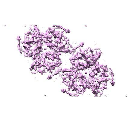 emd_8084.png emd_8084.png | 110.7 KB | ||
| Filedesc structureFactors | emd_8084_sf.cif.gz | 92 KB | ||
| Archive directory |  http://ftp.pdbj.org/pub/emdb/structures/EMD-8084ftp://ftp.pdbj.org/pub/emdb/structures/EMD-8084 http://ftp.pdbj.org/pub/emdb/structures/EMD-8084ftp://ftp.pdbj.org/pub/emdb/structures/EMD-8084 | HTTPS FTP |
-Related structure data
| Related structure data |  5ia9MC  8076C  5i9kC M: atomic model generated by this map C: citing same article ( |
|---|---|
| Similar structure data |
-Links
| EMDB pages | EMDB (EBI/PDBe) / EMDataResource |
|---|---|
| Related items in Molecule of the Month |


-Map
| File | Download / File: emd_8084.map.gz / Format: CCP4 / Size: 1.7 MB / Type: IMAGE STORED AS FLOATING POINT NUMBER (4 BYTES) | ||||||||||||||||||||||||||||||||||||||||||||||||||||||||||||||||||||
|---|---|---|---|---|---|---|---|---|---|---|---|---|---|---|---|---|---|---|---|---|---|---|---|---|---|---|---|---|---|---|---|---|---|---|---|---|---|---|---|---|---|---|---|---|---|---|---|---|---|---|---|---|---|---|---|---|---|---|---|---|---|---|---|---|---|---|---|---|---|
| Annotation | None | ||||||||||||||||||||||||||||||||||||||||||||||||||||||||||||||||||||
| Projections & slices | Image control
Images are generated by Spider. generated in cubic-lattice coordinate | ||||||||||||||||||||||||||||||||||||||||||||||||||||||||||||||||||||
| Voxel size | X: 1.1361 Å / Y: 1.1361 Å / Z: 1.1905 Å | ||||||||||||||||||||||||||||||||||||||||||||||||||||||||||||||||||||
| Density |
| ||||||||||||||||||||||||||||||||||||||||||||||||||||||||||||||||||||
| Symmetry | Space group: 168 | ||||||||||||||||||||||||||||||||||||||||||||||||||||||||||||||||||||
| Details | EMDB XML:
CCP4 map header:
| ||||||||||||||||||||||||||||||||||||||||||||||||||||||||||||||||||||
 Z (Sec.)
 X (Row.)
 Y (Col.)


-Supplemental data
- Sample components
-Entire : The structure of microsomal glutathione transferase 1 in complex ...
| Entire | Name: The structure of microsomal glutathione transferase 1 in complex with the Meisenheimer complex |
|---|---|
| Components |
|
-Supramolecule #1: The structure of microsomal glutathione transferase 1 in complex ...
| Supramolecule | Name: The structure of microsomal glutathione transferase 1 in complex with the Meisenheimer complex type: complex / ID: 1 / Parent: 0 / Macromolecule list: #1 |
|---|---|
| Source (natural) | Organism: Rattus norvegicus (Norway rat) |
| Recombinant expression | Organism:  Escherichia coli (E. coli) / Recombinant plasmid: pSP19T7LT Escherichia coli (E. coli) / Recombinant plasmid: pSP19T7LT |
| Molecular weight | Theoretical: 543.57 KDa |
-Macromolecule #1: Microsomal glutathione S-transferase 1
| Macromolecule | Name: Microsomal glutathione S-transferase 1 / type: protein_or_peptide / ID: 1 / Number of copies: 1 / Enantiomer: LEVO / EC number: glutathione transferase |
|---|---|
| Source (natural) | Organism: Rattus norvegicus (Norway rat) |
| Molecular weight | Theoretical: 17.492488 KDa |
| Recombinant expression | Organism: Escherichia coli (E. coli) |
| Sequence | String: MADLKQLMDN EVLMAFTSYA TIILAKMMFL SSATAFQRLT NKVFANPEDC AGFGKGENAK KFLRTDEKVE RVRRAHLNDL ENIVPFLGI GLLYSLSGPD LSTALIHFRI FVGARIYHTI AYLTPLPQPN RGLAFFVGYG VTLSMAYRLL RSRLYL |
-Macromolecule #2: 1-(S-GLUTATHIONYL)-2,4,6-TRINITROCYCLOHEXA-2,5-DIENE
| Macromolecule | Name: 1-(S-GLUTATHIONYL)-2,4,6-TRINITROCYCLOHEXA-2,5-DIENE / type: ligand / ID: 2 / Number of copies: 1 / Formula: GTD |
|---|---|
| Molecular weight | Theoretical: 520.428 Da |
| Chemical component information |  ChemComp-GTD: |
-Macromolecule #3: 1,2-DIACYL-SN-GLYCERO-3-PHOSPHOCHOLINE
| Macromolecule | Name: 1,2-DIACYL-SN-GLYCERO-3-PHOSPHOCHOLINE / type: ligand / ID: 3 / Number of copies: 2 / Formula: PC1 |
|---|---|
| Molecular weight | Theoretical: 790.145 Da |
| Chemical component information |  ChemComp-PC1: |
-Macromolecule #4: PALMITIC ACID
| Macromolecule | Name: PALMITIC ACID / type: ligand / ID: 4 / Number of copies: 2 / Formula: PLM |
|---|---|
| Molecular weight | Theoretical: 256.424 Da |
| Chemical component information |  ChemComp-PLM: |
-Experimental details
-Structure determination
| Method | cryo EM |
|---|---|
 Processing Processing | electron crystallography |
| Aggregation state | 2D array |
-Sample preparation
| Buffer | pH: 7.4 |
|---|---|
| Sugar embedding | Material: trehalose |
| Vitrification | Cryogen name: NITROGEN |
| Crystal formation | Lipid protein ratio: 3 / Lipid mixture: bovine liver lecithin / Temperature: 303.0 K / Details: dialysis |
- Electron microscopy
| Microscope | JEOL 2100F |
|---|---|
| Electron beam | Acceleration voltage: 200 kV / Electron source: FIELD EMISSION GUN |
| Electron optics | Illumination mode: FLOOD BEAM / Imaging mode: DIFFRACTION / Camera length: 200 mm |
| Image recording | Film or detector model: TVIPS TEMCAM-F415 (4k x 4k) / Average electron dose: 1.0 e/Å2 |
-Image processing
| Crystal parameters | Unit cell - A: 81.8 Å / Unit cell - B: 81.8 Å / Unit cell - C: 100.0 Å / Unit cell - γ: 120.0 ° / Plane group: P 6 |
|---|---|
| Crystallography statistics | Number intensities measured: 43603 / Number structure factors: 3063 / Fourier space coverage: 72.4 / R sym: 12 / R merge: 34.3 / Overall phase error: 0.0001 / Overall phase residual: 0.0001 / Phase error rejection criteria: 0 / High resolution: 3.5 Å |
| Final reconstruction | Resolution.type: BY AUTHOR / Resolution: 3.5 Å / Resolution method: DIFFRACTION PATTERN/LAYERLINES |
