 Movie
Movie Controller

[English] 日本語
 Yorodumi
- EMDB-6683: The 7 angstrom resolution CryoEM map of the bacterial flagellar p... -
+ Open data

- Basic information
| Entry | Database: EMDB / ID: EMD-6683 | |||||||||
|---|---|---|---|---|---|---|---|---|---|---|
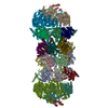
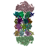
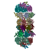
| Title | The 7 angstrom resolution CryoEM map of the bacterial flagellar polyrod | |||||||||
 Map data Map data | ||||||||||
 Sample Sample |
| |||||||||
| Function / homology |  Function and homology information Function and homology informationbacterial-type flagellum basal body, distal rod / bacterial-type flagellum-dependent swarming motility / bacterial-type flagellum-dependent cell motility Similarity search - Function | |||||||||
| Biological species |   Salmonella typhimurium (strain LT2 / SGSC1412 / ATCC 700720) (bacteria) Salmonella typhimurium (strain LT2 / SGSC1412 / ATCC 700720) (bacteria) | |||||||||
| Method | helical reconstruction / cryo EM / Resolution: 7.4 Å | |||||||||
 Authors Authors | Fujii T / Namba K | |||||||||
 Citation Citation | Journal: Nat Commun / Year: 2017 Title: Identical folds used for distinct mechanical functions of the bacterial flagellar rod and hook. Authors: Takashi Fujii / Takayuki Kato / Koichi D Hiraoka / Tomoko Miyata / Tohru Minamino / Fabienne F V Chevance / Kelly T Hughes / Keiichi Namba /   Abstract: The bacterial flagellum is a motile organelle driven by a rotary motor, and its axial portions function as a drive shaft (rod), a universal joint (hook) and a helical propeller (filament). The rod ...The bacterial flagellum is a motile organelle driven by a rotary motor, and its axial portions function as a drive shaft (rod), a universal joint (hook) and a helical propeller (filament). The rod and hook are directly connected to each other, with their subunit proteins FlgG and FlgE having 39% sequence identity, but show distinct mechanical properties; the rod is straight and rigid as a drive shaft whereas the hook is flexible in bending as a universal joint. Here we report the structure of the rod and comparison with that of the hook. While these two structures have the same helical symmetry and repeat distance and nearly identical folds of corresponding domains, the domain orientations differ by ∼7°, resulting in tight and loose axial subunit packing in the rod and hook, respectively, conferring the rigidity on the rod and flexibility on the hook. This provides a good example of versatile use of a protein structure in biological organisms. | |||||||||
| History |
|
- Structure visualization
| Movie |
Movie viewer |
|---|---|
| Structure viewer | EM map: SurfViewMolmilJmol/JSmol |
| Supplemental images |

- Downloads & links
-EMDB archive
| Map data | emd_6683.map.gz | 3 MB | EMDB map data format | |
|---|---|---|---|---|
| Header (meta data) | emd-6683-v30.xmlemd-6683.xml | 8.9 KB 8.9 KB | Display Display | EMDB header |
| Images | 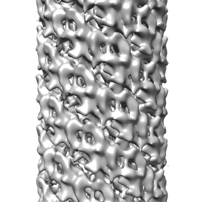 emd_6683.png emd_6683.png | 90.4 KB | ||
| Archive directory |  http://ftp.pdbj.org/pub/emdb/structures/EMD-6683ftp://ftp.pdbj.org/pub/emdb/structures/EMD-6683 http://ftp.pdbj.org/pub/emdb/structures/EMD-6683ftp://ftp.pdbj.org/pub/emdb/structures/EMD-6683 | HTTPS FTP |
-Related structure data
| Related structure data |  5wrhMC  6jzrM M: atomic model generated by this map C: citing same article ( |
|---|---|
| Similar structure data |


-Links
| EMDB pages | EMDB (EBI/PDBe) / EMDataResource |
|---|---|
| Related items in Molecule of the Month |

-Map
| File | Download / File: emd_6683.map.gz / Format: CCP4 / Size: 12.9 MB / Type: IMAGE STORED AS FLOATING POINT NUMBER (4 BYTES) | ||||||||||||||||||||||||||||||||||||||||||||||||||||||||||||
|---|---|---|---|---|---|---|---|---|---|---|---|---|---|---|---|---|---|---|---|---|---|---|---|---|---|---|---|---|---|---|---|---|---|---|---|---|---|---|---|---|---|---|---|---|---|---|---|---|---|---|---|---|---|---|---|---|---|---|---|---|---|
| Voxel size | X=Y=Z: 1.64 Å | ||||||||||||||||||||||||||||||||||||||||||||||||||||||||||||
| Density |
| ||||||||||||||||||||||||||||||||||||||||||||||||||||||||||||
| Symmetry | Space group: 1 | ||||||||||||||||||||||||||||||||||||||||||||||||||||||||||||
| Details | EMDB XML:
CCP4 map header:
| ||||||||||||||||||||||||||||||||||||||||||||||||||||||||||||
-Supplemental data
- Sample components
-Entire : the bacterial flagellar polyrod
| Entire | Name: the bacterial flagellar polyrod |
|---|---|
| Components |
|
-Supramolecule #1: the bacterial flagellar polyrod
| Supramolecule | Name: the bacterial flagellar polyrod / type: complex / ID: 1 / Parent: 0 / Macromolecule list: all |
|---|---|
| Source (natural) | Organism: Salmonella typhimurium (strain LT2 / SGSC1412 / ATCC 700720) (bacteria) |
-Macromolecule #1: Flagellar basal-body rod protein FlgG
| Macromolecule | Name: Flagellar basal-body rod protein FlgG / type: protein_or_peptide / ID: 1 / Number of copies: 1 / Enantiomer: LEVO |
|---|---|
| Source (natural) | Organism: Salmonella typhimurium (strain LT2 / SGSC1412 / ATCC 700720) (bacteria) Strain: LT2 / SGSC1412 / ATCC 700720 |
| Molecular weight | Theoretical: 27.784807 KDa |
| Sequence | String: MISSLWIAKT GLDAQQTNMD VIANNLANVS TNGFKRQRAV FEDLLYQTIR QPGAQSSEQT TLPSGLQIGT GVRPVATERL HSQGNLSQT NNSKDVAIKG QGFFQVMLPD GTSAYTRDGS FQVDQNGQLV TAGGFQVQPA ITIPANALSI TIGRDGVVSV T QQGQAAPV ...String: MISSLWIAKT GLDAQQTNMD VIANNLANVS TNGFKRQRAV FEDLLYQTIR QPGAQSSEQT TLPSGLQIGT GVRPVATERL HSQGNLSQT NNSKDVAIKG QGFFQVMLPD GTSAYTRDGS FQVDQNGQLV TAGGFQVQPA ITIPANALSI TIGRDGVVSV T QQGQAAPV QVGQLNLTTF MNDTGLESIG ENLYIETQSS GAPNESTPGL NGAGLLYQGY VETSNVNVAE ELVNMIQVQR AY EINSKAV STTDQMLQKL TQL |
-Experimental details
-Structure determination
| Method | cryo EM |
|---|---|
 Processing Processing | helical reconstruction |
| Aggregation state | filament |
-Sample preparation
| Buffer | pH: 8 |
|---|---|
| Vitrification | Cryogen name: ETHANE |
- Electron microscopy
| Microscope | JEOL 3200FSC |
|---|---|
| Electron beam | Acceleration voltage: 200 kV / Electron source: FIELD EMISSION GUN |
| Electron optics | Illumination mode: FLOOD BEAM / Imaging mode: BRIGHT FIELDBright-field microscopy |
| Image recording | Film or detector model: TVIPS TEMCAM-F415 (4k x 4k) / Average electron dose: 20.0 e/Å2 |
-Image processing
| Startup model | Type of model: OTHER / Details: simple cylinder |
|---|---|
| Final angle assignment | Type: NOT APPLICABLE |
| Final reconstruction | Applied symmetry - Helical parameters - Δz: 4.13 Å Applied symmetry - Helical parameters - Δ&Phi: 64.75 ° Applied symmetry - Helical parameters - Axial symmetry: C1 (asymmetric) Resolution.type: BY AUTHOR / Resolution: 7.4 Å / Resolution method: FSC 0.5 CUT-OFF / Number images used: 10645 |
