Movie
Movie Controller


+ Open data

- Basic information
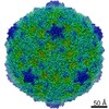
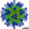
| Entry | Database: EMDB / ID: EMD-6453 | |||||||||
|---|---|---|---|---|---|---|---|---|---|---|
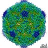
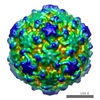
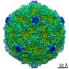
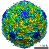
| Title | Electron cryo-microscopy of a betanodavirus virus-like particle | |||||||||
 Map data Map data | Reconstruction of virus-like particle orange-spotted grouper nervous necrosis virus (OGNNV) | |||||||||
 Sample Sample |
| |||||||||
 Keywords Keywords |  virus-like particle / orange-spotted grouper nervous necrosis virus / betanodavirus virus-like particle / orange-spotted grouper nervous necrosis virus / betanodavirus | |||||||||
| Function / homology | Nodavirus capsid / nodavirus capsid protein / Viral coat protein subunit / Virus-like particle of orange-spotted grouper nervous necrosis virus Function and homology information Function and homology information | |||||||||
| Biological species |  Orange-spotted grouper nervous necrosis virus Orange-spotted grouper nervous necrosis virus | |||||||||
| Method | single particle reconstruction / cryo EM / Resolution: 3.9 Å | |||||||||
 Authors Authors | Junfeng XIE / Kunpeng LI / Yuanzhu GAO / Runqing HUANG / Yuxiong LAI / Yan SHI / Shaowei YANG / Guohua ZHU / Qinfen ZHANG / Jianguo HE | |||||||||
 Citation Citation | Journal: Vet Res / Year: 2016 Title: Structural analysis and insertion study reveal the ideal sites for surface displaying foreign peptides on a betanodavirus-like particle. Authors: Junfeng Xie / Kunpeng Li / Yuanzhu Gao / Runqing Huang / Yuxiong Lai / Yan Shi / Shaowei Yang / Guohua Zhu / Qinfen Zhang / Jianguo He /  Abstract: Betanodavirus infection causes fatal disease of viral nervous necrosis in many cultured marine and freshwater fish worldwide and the virus-like particles (VLP) are effective vaccines against ...Betanodavirus infection causes fatal disease of viral nervous necrosis in many cultured marine and freshwater fish worldwide and the virus-like particles (VLP) are effective vaccines against betanodavirus. But vaccine and viral vector designs of betanodavirus VLP based on their structures remain lacking. Here, the three-dimensional structure of orange-spotted grouper nervous necrosis virus (OGNNV) VLP (RBS) at 3.9 Å reveals the organization of capsid proteins (CP). Based on the structural results, seven putative important sites were selected to genetically insert a 6× histidine (His)-tag for VLP formation screen, resulting in four His-tagged VLP (HV) at positions N-terminus, Ala220, Pro292 and C-terminus. The His-tags of N-terminal HV (NHV) were concealed inside virions while those of 220HV and C-terminal HV (CHV) were displayed at the outer surface. NHV, 220HV and CHV maintained the same cell entry ability as RBS in the Asian sea bass (SB) cell line, indicating that their similar surface structures can be recognized by the cellular entry receptor(s). For application of vaccine design, chromatography-purified CHV could provoke NNV-specific antibody responses as strong as those of RBS in a sea bass immunization assay. Furthermore, in carrying capacity assays, N-terminus and Ala220 can only carry short peptides and C-terminus can even accommodate large protein such as GFP to generate fluorescent VLP (CGV). For application of a viral vector, CGV could be real-time visualized to enter SB cells in invasion study. All the results confirmed that the C-terminus of CP is a suitable site to accommodate foreign peptides for vaccine design and viral vector development. | |||||||||
| History |
|
- Structure visualization
| Movie |
Movie viewer |
|---|---|
| Structure viewer | EM map: SurfViewMolmilJmol/JSmol |
| Supplemental images |

- Downloads & links
-EMDB archive
| Map data | emd_6453.map.gz | 328.2 MB | EMDB map data format | |
|---|---|---|---|---|
| Header (meta data) | emd-6453-v30.xmlemd-6453.xml | 11.2 KB 11.2 KB | Display Display | EMDB header |
| Images | 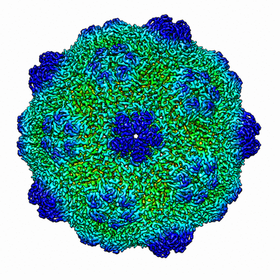 400_6453.gif 400_6453.gif 80_6453.gif 80_6453.gif | 109.4 KB 5.6 KB | ||
| Archive directory |  http://ftp.pdbj.org/pub/emdb/structures/EMD-6453ftp://ftp.pdbj.org/pub/emdb/structures/EMD-6453 http://ftp.pdbj.org/pub/emdb/structures/EMD-6453ftp://ftp.pdbj.org/pub/emdb/structures/EMD-6453 | HTTPS FTP |
-Related structure data
| Related structure data |  3jbmMC M: atomic model generated by this map C: citing same article ( |
|---|---|
| Similar structure data |


-Links
| EMDB pages | EMDB (EBI/PDBe) / EMDataResource |
|---|
-Map
| File | Download / File: emd_6453.map.gz / Format: CCP4 / Size: 412 MB / Type: IMAGE STORED AS FLOATING POINT NUMBER (4 BYTES) | ||||||||||||||||||||||||||||||||||||||||||||||||||||||||||||
|---|---|---|---|---|---|---|---|---|---|---|---|---|---|---|---|---|---|---|---|---|---|---|---|---|---|---|---|---|---|---|---|---|---|---|---|---|---|---|---|---|---|---|---|---|---|---|---|---|---|---|---|---|---|---|---|---|---|---|---|---|---|
| Annotation | Reconstruction of virus-like particle orange-spotted grouper nervous necrosis virus (OGNNV) | ||||||||||||||||||||||||||||||||||||||||||||||||||||||||||||
| Voxel size | X=Y=Z: 0.933 Å | ||||||||||||||||||||||||||||||||||||||||||||||||||||||||||||
| Density |
| ||||||||||||||||||||||||||||||||||||||||||||||||||||||||||||
| Symmetry | Space group: 1 | ||||||||||||||||||||||||||||||||||||||||||||||||||||||||||||
| Details | EMDB XML:
CCP4 map header:
| ||||||||||||||||||||||||||||||||||||||||||||||||||||||||||||
-Supplemental data
- Sample components
-Entire : Virus-like particle of orange-spotted grouper nervous necrosis vi...
| Entire | Name: Virus-like particle of orange-spotted grouper nervous necrosis virus generated in E.coli |
|---|---|
| Components |
|
-Supramolecule #1000: Virus-like particle of orange-spotted grouper nervous necrosis vi...
| Supramolecule | Name: Virus-like particle of orange-spotted grouper nervous necrosis virus generated in E.coli type: sample / ID: 1000 / Number unique components: 1 |
|---|---|
| Molecular weight | Theoretical: 6.8 MDa |
-Supramolecule #1: Orange-spotted grouper nervous necrosis virus
| Supramolecule | Name: Orange-spotted grouper nervous necrosis virus / type: virus / ID: 1 / NCBI-ID: 421750 Sci species name: Orange-spotted grouper nervous necrosis virus Virus type: VIRUS-LIKE PARTICLE / Virus isolate: SPECIES / Virus enveloped: No / Virus empty: No |
|---|---|
| Host (natural) | Organism:  Epinephelus coioides (orange-spotted grouper) / synonym: VERTEBRATES Epinephelus coioides (orange-spotted grouper) / synonym: VERTEBRATES |
| Host system | Organism:  Escherichia coli (E. coli) / Recombinant strain: M15 / Recombinant plasmid: pQE30 Escherichia coli (E. coli) / Recombinant strain: M15 / Recombinant plasmid: pQE30 |
| Molecular weight | Theoretical: 6.8 MDa |
| Virus shell | Shell ID: 1 / Diameter: 380 Å / T number (triangulation number): 3 |
-Experimental details
-Structure determination
| Method | cryo EM |
|---|---|
 Processing Processing | single particle reconstruction |
| Aggregation state | particle |
-Sample preparation
| Concentration | 1.5 mg/mL |
|---|---|
| Buffer | pH: 8 / Details: PBS buffer |
| Grid | Details: 300 mesh Quantifoil copper grid with thin carbon support, glow discharged in air atmosphere |
| Vitrification | Cryogen name: ETHANE / Chamber humidity: 100 % / Chamber temperature: 100 K / Instrument: FEI VITROBOT MARK IV / Method: blot for 5 min before plunging |
- Electron microscopy
| Microscope | FEI TITAN KRIOS |
|---|---|
| Electron beam | Acceleration voltage: 300 kV / Electron source: FIELD EMISSION GUN |
| Electron optics | Illumination mode: FLOOD BEAM / Imaging mode: BRIGHT FIELDBright-field microscopy / Cs: 2.7 mm / Nominal defocus max: 2.91 µm / Nominal defocus min: 1.0 µm / Nominal magnification: 96000 |
| Sample stage | Specimen holder model: FEI TITAN KRIOS AUTOGRID HOLDER |
| Temperature | Min: 95 K / Max: 105 K / Average: 100 K |
| Alignment procedure | Legacy - Astigmatism: Objective lens astigmatism was corrected at 200000 times magnification |
| Details | Weak beam illumination |
| Date | Feb 26, 2011 |
| Image recording | Category: CCD / Film or detector model: GATAN ULTRASCAN 4000 (4k x 4k) / Digitization - Sampling interval: 2.5 µm / Number real images: 1359 / Average electron dose: 20 e/Å2 / Bits/pixel: 16 |
| Experimental equipment |  Model: Titan Krios / Image courtesy: FEI Company |
-Image processing
| CTF correction | Details: Each particle |
|---|---|
| Final reconstruction | Algorithm: OTHER / Resolution.type: BY AUTHOR / Resolution: 3.9 Å / Resolution method: OTHER / Software - Name: EMAN, JSPR / Number images used: 32000 |
| Details | The particles were selected manually. |