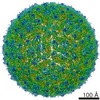
Journal: J Virol / Year: 2015 Title: Three-dimensional structure of a protozoal double-stranded RNA virus that infects the enteric pathogen Giardia lamblia. Authors: Mandy E W Janssen / Yuko Takagi / Kristin N Parent / Giovanni Cardone / Max L Nibert / Timothy S Baker / Abstract: Giardia lamblia virus (GLV) is a small, nonenveloped, nonsegmented double-stranded RNA (dsRNA) virus infecting Giardia lamblia, the most common protozoan pathogen of the human intestine and a major ...Giardia lamblia virus (GLV) is a small, nonenveloped, nonsegmented double-stranded RNA (dsRNA) virus infecting Giardia lamblia, the most common protozoan pathogen of the human intestine and a major agent of waterborne diarrheal disease worldwide. GLV (genus Giardiavirus) is a member of family Totiviridae, along with several other groups of protozoal or fungal viruses, including Leishmania RNA viruses and Trichomonas vaginalis viruses. Interestingly, GLV is more closely related than other Totiviridae members to a group of recently discovered metazoan viruses that includes penaeid shrimp infectious myonecrosis virus (IMNV). Moreover, GLV is the only known protozoal dsRNA virus that can transmit efficiently by extracellular means, also like IMNV. In this study, we used transmission electron cryomicroscopy and icosahedral image reconstruction to examine the GLV virion at an estimated resolution of 6.0 Å. Its outermost diameter is 485 Å, making it the largest totivirus capsid analyzed to date. Structural comparisons of GLV and other totiviruses highlighted a related "T=2" capsid organization and a conserved helix-rich fold in the capsid subunits. In agreement with its unique capacity as a protozoal dsRNA virus to survive and transmit through extracellular environments, GLV was found to be more thermoresistant than Trichomonas vaginalis virus 1, but no specific protein machinery to mediate cell entry, such as the fiber complexes in IMNV, could be localized. These and other structural and biochemical findings provide a basis for future work to dissect the cell entry mechanism of GLV into a "primitive" (early-branching) eukaryotic host and an important enteric pathogen of humans. IMPORTANCE: Numerous pathogenic bacteria, including Corynebacterium diphtheriae, Salmonella enterica, and Vibrio cholerae, are infected with lysogenic bacteriophages that contribute significantly to ...IMPORTANCE: Numerous pathogenic bacteria, including Corynebacterium diphtheriae, Salmonella enterica, and Vibrio cholerae, are infected with lysogenic bacteriophages that contribute significantly to bacterial virulence. In line with this phenomenon, several pathogenic protozoa, including Giardia lamblia, Leishmania species, and Trichomonas vaginalis are persistently infected with dsRNA viruses, and growing evidence indicates that at least some of these protozoal viruses can likewise enhance the pathogenicity of their hosts. Understanding of these protozoal viruses, however, lags far behind that of many bacteriophages. Here, we investigated the dsRNA virus that infects the widespread enteric parasite Giardia lamblia. Using electron cryomicroscopy and icosahedral image reconstruction, we determined the virion structure of Giardia lamblia virus, obtaining new information relating to its assembly, stability, functions in cell entry and transcription, and similarities and differences with other dsRNA viruses. The results of our study set the stage for further mechanistic work on the roles of these viruses in protozoal virulence.
History
Deposition
Apr 16, 2014
-
Header (metadata) release
Apr 30, 2014
-
Map release
Nov 12, 2014
-
Update
May 27, 2015
-
Current status
May 27, 2015
Processing site: RCSB / Status: Released
-
Structure visualization
Movie
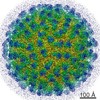
Surface view with section colored by density value
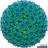
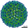
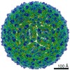
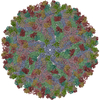
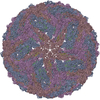
Shell ID: 1 / Diameter: 485 Å / T number (triangulation number): 2
-
Experimental details
-
Structure determination
Method
cryo EM
Processing
single particle reconstruction
Aggregation state
particle
-
Sample preparation
Concentration
0.02 mg/mL
Buffer
pH: 7.2 / Details: 50 mM HEPES, 500 mM NaCl, 20 mM MgCl2
Grid
Details: Quantifoil R2/2
Vitrification
Cryogen name: ETHANE / Chamber humidity: 99 % / Chamber temperature: 90 K / Instrument: HOMEMADE PLUNGER / Method: Blot 5 sec before plunging
-
Electron microscopy
Microscope
FEI POLARA 300
Temperature
Min: 90 K / Max: 96 K / Average: 93 K
Alignment procedure
Legacy - Astigmatism: Objective lens astigmatism was corrected at 135,000 times magnification.
Date
Apr 18, 2012
Image recording
Category: FILM / Film or detector model: KODAK SO-163 FILM / Digitization - Scanner: OTHER / Digitization - Sampling interval: 6.35 µm / Number real images: 101 / Average electron dose: 25 e/Å2 / Bits/pixel: 8
Tilt angle min
0
Tilt angle max
0
Electron beam
Acceleration voltage: 200 kV / Electron source: FIELD EMISSION GUN
Model: Tecnai Polara / Image courtesy: FEI Company
-
Image processing
Details
Particles were selected and preprocessed using RobEM. Image reconstruction was performed using Auto3DEM.
CTF correction
Details: robem
Final reconstruction
Algorithm: OTHER / Resolution.type: BY AUTHOR / Resolution: 6.0 Å / Resolution method: OTHER / Software - Name: auto3dem Details: Particles were selected and preprocessed using RobEM. Number images used: 14125
+
About Yorodumi
-
News
-
Feb 9, 2022. New format data for meta-information of EMDB entries
New format data for meta-information of EMDB entries
Version 3 of the EMDB header file is now the official format.
The previous official version 1.9 will be removed from the archive.
In the structure databanks used in Yorodumi, some data are registered as the other names, "COVID-19 virus" and "2019-nCoV". Here are the details of the virus and the list of structure data.
Jan 31, 2019. EMDB accession codes are about to change! (news from PDBe EMDB page)
EMDB accession codes are about to change! (news from PDBe EMDB page)
The allocation of 4 digits for EMDB accession codes will soon come to an end. Whilst these codes will remain in use, new EMDB accession codes will include an additional digit and will expand incrementally as the available range of codes is exhausted. The current 4-digit format prefixed with “EMD-” (i.e. EMD-XXXX) will advance to a 5-digit format (i.e. EMD-XXXXX), and so on. It is currently estimated that the 4-digit codes will be depleted around Spring 2019, at which point the 5-digit format will come into force.
The EM Navigator/Yorodumi systems omit the EMD- prefix.
Related info.:Q: What is EMD? / ID/Accession-code notation in Yorodumi/EM Navigator
Yorodumi is a browser for structure data from EMDB, PDB, SASBDB, etc.
This page is also the successor to EM Navigator detail page, and also detail information page/front-end page for Omokage search.
The word "yorodu" (or yorozu) is an old Japanese word meaning "ten thousand". "mi" (miru) is to see.
Related info.:EMDB / PDB / SASBDB / Comparison of 3 databanks / Yorodumi Search / Aug 31, 2016. New EM Navigator & Yorodumi / Yorodumi Papers / Jmol/JSmol / Function and homology information / Changes in new EM Navigator and Yorodumi
 Movie
Movie Controller

 Yorodumi
 Open data
 Basic information
 Map data
 Sample
 Keywords
 Giardia lamblia virus
 Authors
 Citation
 Structure visualization
 Movie viewer
 Downloads & links
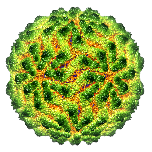 emd_5948.png
 http://ftp.pdbj.org/pub/emdb/structures/EMD-5948
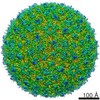


 Sample components
 Giardia intestinalis (eukaryote) / Strain: WBI / synonym: PROTOZOA
 Processing
 Electron microscopy
 FIELD EMISSION GUN
