 Movie
Movie Controller


+ Open data

- Basic information
| Entry | Database: EMDB / ID: EMD-5383 | |||||||||
|---|---|---|---|---|---|---|---|---|---|---|
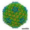
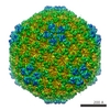
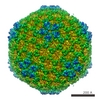
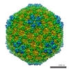
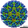
| Title | Marine Bacteriophage SIO-2 with T12 symmetry (Procapsid) | |||||||||
 Map data Map data | SIO-2 prophage | |||||||||
 Sample Sample |
| |||||||||
 Keywords Keywords | procapsid / Marine Bacteriophage / Phage / Virus / Decoration Proteins / Ig-like | |||||||||
| Biological species |  Vibrio phage SIO-2 (virus) Vibrio phage SIO-2 (virus) | |||||||||
| Method | single particle reconstruction / cryo EM / Resolution: 15.0 Å | |||||||||
 Authors Authors | Lander GC / Baudoux AC / Azam F / Potter CS / Carragher B / Johnson JE | |||||||||
 Citation Citation | Journal: Structure / Year: 2012 Title: Capsomer dynamics and stabilization in the T = 12 marine bacteriophage SIO-2 and its procapsid studied by CryoEM. Authors: Gabriel C Lander / Anne-Claire Baudoux / Farooq Azam / Clinton S Potter / Bridget Carragher / John E Johnson /  Abstract: We report the subnanometer cryo-electron microscopy (cryoEM) reconstruction of a marine siphovirus, the Vibrio phage SIO-2. This phage is lytic for related Vibrio species with significant ecological ...We report the subnanometer cryo-electron microscopy (cryoEM) reconstruction of a marine siphovirus, the Vibrio phage SIO-2. This phage is lytic for related Vibrio species with significant ecological importance, including the broadly antagonistic bacterium Vibrio sp. SWAT3. The three-dimensional structure of the 800 Å SIO-2, icosahedrally averaged head of the tailed particle revealed a T = 12 quasi-symmetry not previously described in a bacteriophage. Two morphologically distinct types of auxiliary proteins were also identified; one species bound to the surface of hexamers, and the other bound to pentamers. The secondary structure, evident in the electron density, shows that the major capsid protein has the HK97-like fold. The three-dimensional structure of the procapsid form, also presented here, has no "decoration" proteins and reveals a capsomer organization due to the constraints of the T = 12 symmetry. | |||||||||
| History |
|
- Structure visualization
| Movie |
 Movie viewer Movie viewer |
|---|---|
| Structure viewer | EM map: SurfViewMolmilJmol/JSmol |
| Supplemental images |

- Downloads & links
-EMDB archive
| Map data | emd_5383.map.gz | 10.2 MB | EMDB map data format | |
|---|---|---|---|---|
| Header (meta data) | emd-5383-v30.xmlemd-5383.xml | 10.1 KB 10.1 KB | Display Display | EMDB header |
| Images | 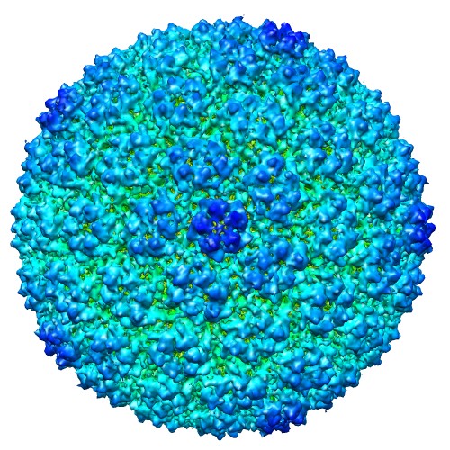 emd_5383_1.jpg emd_5383_1.jpg | 111.6 KB | ||
| Archive directory |  http://ftp.pdbj.org/pub/emdb/structures/EMD-5383ftp://ftp.pdbj.org/pub/emdb/structures/EMD-5383 http://ftp.pdbj.org/pub/emdb/structures/EMD-5383ftp://ftp.pdbj.org/pub/emdb/structures/EMD-5383 | HTTPS FTP |
-Validation report
| Summary document | emd_5383_validation.pdf.gz | 78.3 KB | Display | EMDB validaton report |
|---|---|---|---|---|
| Full document | emd_5383_full_validation.pdf.gz | 77.4 KB | Display | |
| Data in XML | emd_5383_validation.xml.gz | 493 B | Display | |
| Arichive directory | https://ftp.pdbj.org/pub/emdb/validation_reports/EMD-5383ftp://ftp.pdbj.org/pub/emdb/validation_reports/EMD-5383 | HTTPS FTP |
-Related structure data

-Links
| EMDB pages | EMDB (EBI/PDBe) / EMDataResource |
|---|
-Map
| File | Download / File: emd_5383.map.gz / Format: CCP4 / Size: 21.7 MB / Type: IMAGE STORED AS FLOATING POINT NUMBER (4 BYTES) | ||||||||||||||||||||||||||||||||||||||||||||||||||||||||||||||||||||
|---|---|---|---|---|---|---|---|---|---|---|---|---|---|---|---|---|---|---|---|---|---|---|---|---|---|---|---|---|---|---|---|---|---|---|---|---|---|---|---|---|---|---|---|---|---|---|---|---|---|---|---|---|---|---|---|---|---|---|---|---|---|---|---|---|---|---|---|---|---|
| Annotation | SIO-2 prophage | ||||||||||||||||||||||||||||||||||||||||||||||||||||||||||||||||||||
| Voxel size | X=Y=Z: 3.9 Å | ||||||||||||||||||||||||||||||||||||||||||||||||||||||||||||||||||||
| Density |
| ||||||||||||||||||||||||||||||||||||||||||||||||||||||||||||||||||||
| Symmetry | Space group: 1 | ||||||||||||||||||||||||||||||||||||||||||||||||||||||||||||||||||||
| Details | EMDB XML:
CCP4 map header:
| ||||||||||||||||||||||||||||||||||||||||||||||||||||||||||||||||||||
-Supplemental data
- Sample components
-Entire : Marine Phage SIO-2
| Entire | Name: Marine Phage SIO-2 |
|---|---|
| Components |
|
-Supramolecule #1000: Marine Phage SIO-2
| Supramolecule | Name: Marine Phage SIO-2 / type: sample / ID: 1000 / Details: procapsid / Oligomeric state: immature capsid / Number unique components: 1 |
|---|---|
| Molecular weight | Theoretical: 12.2 MDa |
-Supramolecule #1: Vibrio phage SIO-2
| Supramolecule | Name: Vibrio phage SIO-2 / type: virus / ID: 1 / Name.synonym: SIO-2 / Details: particles were present with mature phage prep / NCBI-ID: 700512 / Sci species name: Vibrio phage SIO-2 / Database: NCBI / Virus type: VIRION / Virus isolate: STRAIN / Virus enveloped: No / Virus empty: Yes / Syn species name: SIO-2 |
|---|---|
| Host (natural) | Organism: Vibrio sp. SWAT-3 / synonym: BACTERIA(EUBACTERIA) |
| Molecular weight | Theoretical: 12.2 MDa |
| Virus shell | Shell ID: 1 / Name: gene84 / Diameter: 600 Å / T number (triangulation number): 12 |
-Experimental details
-Structure determination
| Method | cryo EM |
|---|---|
 Processing Processing | single particle reconstruction |
| Aggregation state | particle |
-Sample preparation
| Buffer | Details: 100 kDa-filtered autoclaved seawater |
|---|---|
| Grid | Details: 200 mesh Cu grid |
| Vitrification | Cryogen name: ETHANE / Chamber humidity: 100 % / Chamber temperature: 78 K / Instrument: OTHER / Details: Vitrification instrument: Vitrobot / Method: blot for 4 seconds before plunging |
- Electron microscopy
| Microscope | FEI TECNAI 20 |
|---|---|
| Temperature | Min: 78 K / Max: 78 K / Average: 78 K |
| Alignment procedure | Legacy - Astigmatism: objective lens astigmatism was corrected at 210K times magnification |
| Details | Collected using Leginon software |
| Date | Jun 13, 2009 |
| Image recording | Category: CCD / Film or detector model: TVIPS TEMCAM-F415 (4k x 4k) / Digitization - Sampling interval: 0.975 µm / Number real images: 6628 / Average electron dose: 30 e/Å2 |
| Electron beam | Acceleration voltage: 200 kV / Electron source:  FIELD EMISSION GUN FIELD EMISSION GUN |
| Electron optics | Calibrated magnification: 80000 / Illumination mode: FLOOD BEAM / Imaging mode: BRIGHT FIELD / Cs: 2.0 mm / Nominal defocus max: 3.0 µm / Nominal defocus min: 1.8 µm / Nominal magnification: 80000 |
| Sample stage | Specimen holder: Gatan CT3500 / Specimen holder model: GATAN LIQUID NITROGEN |
-Image processing
| Details | particles automatically selected and manually edited |
|---|---|
| CTF correction | Details: whole micrograph |
| Final reconstruction | Algorithm: OTHER / Resolution.type: BY AUTHOR / Resolution: 15.0 Å / Resolution method: FSC 0.5 CUT-OFF / Software - Name: EMAN and Frealign / Number images used: 1179 |
