Journal: PLoS One / Year: 2011 Title: Structural basis for variant-specific neuroligin-binding by α-neurexin. Authors: Hiroki Tanaka / Terukazu Nogi / Norihisa Yasui / Kenji Iwasaki / Junichi Takagi / Abstract: Neurexins (Nrxs) are presynaptic membrane proteins with a single membrane-spanning domain that mediate asymmetric trans-synaptic cell adhesion by binding to their postsynaptic receptor neuroligins. ...Neurexins (Nrxs) are presynaptic membrane proteins with a single membrane-spanning domain that mediate asymmetric trans-synaptic cell adhesion by binding to their postsynaptic receptor neuroligins. α-Nrx has a large extracellular region comprised of multiple copies of laminin, neurexin, sex-hormone-binding globulin (LNS) domains and epidermal growth factor (EGF) modules, while that of β-Nrx has but a single LNS domain. It has long been known that the larger α-Nrx and the shorter β-Nrx show distinct binding behaviors toward different isoforms/variants of neuroligins, although the underlying mechanism has yet to be elucidated. Here, we describe the crystal structure of a fragment corresponding to the C-terminal one-third of the Nrx1α ectodomain, consisting of LNS5-EGF3-LNS6. The 2.3 Å-resolution structure revealed the presence of a domain configuration that was rigidified by inter-domain contacts, as opposed to the more common flexible "beads-on-a-string" arrangement. Although the neuroligin-binding site on the LNS6 domain was completely exposed, the location of the α-Nrx specific LNS5-EGF3 segment proved incompatible with the loop segment inserted in the B+ neuroligin variant, which explains the variant-specific neuroligin recognition capability observed in α-Nrx. This, combined with a low-resolution molecular envelope obtained by a single particle reconstruction performed on negatively stained full-length Nrx1α sample, allowed us to derive a structural model of the α-Nrx ectodomain. This model will help us understand not only how the large α-Nrx ectodomain is accommodated in the synaptic cleft, but also how the trans-synaptic adhesion mediated by α- and β-Nrxs could differentially affect synaptic structure and function.
History
Deposition
Mar 24, 2011
-
Header (metadata) release
Jun 23, 2011
-
Map release
Jun 27, 2011
-
Update
Sep 23, 2011
-
Current status
Sep 23, 2011
Processing site: RCSB / Status: Released
-
Structure visualization
Movie
Surface view with section colored by density value
Download / File: emd_5270.map.gz / Format: CCP4 / Size: 29.8 MB / Type: IMAGE STORED AS FLOATING POINT NUMBER (4 BYTES)
Annotation
This is a 3-D map of the entire ectodomain of bovine Nrx1alpha
Voxel size
X=Y=Z: 2.2 Å
Density
Contour Level
By AUTHOR: 7.59 / Movie #1: 7.59
Minimum - Maximum
-11.7317 - 29.890899999999998
Average (Standard dev.)
-0.0000000071204 (±1.0)
Symmetry
Space group: 1
Details
EMDB XML:
Map geometry
Axis order
X
Y
Z
Origin
-100
-100
-100
Dimensions
200
200
200
Spacing
200
200
200
Cell
A=B=C: 440 Å α=β=γ: 90 °
CCP4 map header:
mode
Image stored as Reals
Å/pix. X/Y/Z
2.2
2.2
2.2
M x/y/z
200
200
200
origin x/y/z
0.000
0.000
0.000
length x/y/z
440.000
440.000
440.000
α/β/γ
90.000
90.000
90.000
start NX/NY/NZ
-62
-62
-62
NX/NY/NZ
125
125
125
MAP C/R/S
1
2
3
start NC/NR/NS
-100
-100
-100
NC/NR/NS
200
200
200
D min/max/mean
-11.732
29.891
-0.000
-
Supplemental data
-
Sample components
-
Entire : an ectodomain fragment of alpha-neurexin 1
Entire
Name: an ectodomain fragment of alpha-neurexin 1
Components
Sample: an ectodomain fragment of alpha-neurexin 1
Protein or peptide: Neurexin
-
Supramolecule #1000: an ectodomain fragment of alpha-neurexin 1
Supramolecule
Name: an ectodomain fragment of alpha-neurexin 1 / type: sample / ID: 1000 Details: The sample was subjected to a size exclusion chromatography before negatively staining Oligomeric state: monomer / Number unique components: 1
In the structure databanks used in Yorodumi, some data are registered as the other names, "COVID-19 virus" and "2019-nCoV". Here are the details of the virus and the list of structure data.
Jan 31, 2019. EMDB accession codes are about to change! (news from PDBe EMDB page)
EMDB accession codes are about to change! (news from PDBe EMDB page)
The allocation of 4 digits for EMDB accession codes will soon come to an end. Whilst these codes will remain in use, new EMDB accession codes will include an additional digit and will expand incrementally as the available range of codes is exhausted. The current 4-digit format prefixed with “EMD-” (i.e. EMD-XXXX) will advance to a 5-digit format (i.e. EMD-XXXXX), and so on. It is currently estimated that the 4-digit codes will be depleted around Spring 2019, at which point the 5-digit format will come into force.
The EM Navigator/Yorodumi systems omit the EMD- prefix.
Related info.:Q: What is EMD? / ID/Accession-code notation in Yorodumi/EM Navigator
Yorodumi is a browser for structure data from EMDB, PDB, SASBDB, etc.
This page is also the successor to EM Navigator detail page, and also detail information page/front-end page for Omokage search.
The word "yorodu" (or yorozu) is an old Japanese word meaning "ten thousand". "mi" (miru) is to see.
Related info.:EMDB / PDB / SASBDB / Comparison of 3 databanks / Yorodumi Search / Aug 31, 2016. New EM Navigator & Yorodumi / Yorodumi Papers / Jmol/JSmol / Function and homology information / Changes in new EM Navigator and Yorodumi
 Movie
Movie Controller


 Open data
 Basic information
 Map data
 Sample
 Keywords
 synapse / neulexin /
  Function and homology information
 Authors
 Citation
 Structure visualization
 Downloads & links
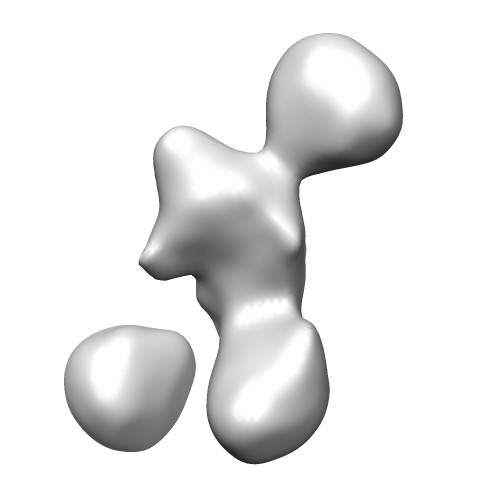 emd_5270_1.jpg
 http://ftp.pdbj.org/pub/emdb/structures/EMD-5270
 Sample components
 Processing
 Electron microscopy