 Movie
Movie Controller


+ Open data

- Basic information
| Entry | Database: EMDB / ID: EMD-5114 | |||||||||
|---|---|---|---|---|---|---|---|---|---|---|
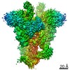
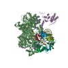
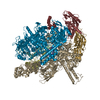
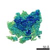
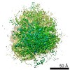
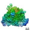
| Title | BK channel with membrane density subtracted | |||||||||
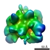
 Map data Map data | Surface rendering of the inside-out BK channel map with membrane density subtracted. The mesh shows a membrane-patch difference map. | |||||||||
 Sample Sample |
| |||||||||
 Keywords Keywords |  Potassium channel Potassium channel | |||||||||
| Biological species |  Homo sapiens (human) Homo sapiens (human) | |||||||||
| Method | single particle reconstruction / cryo EM / Resolution: 17.0 Å | |||||||||
 Authors Authors | Wang L / Sigworth FJ | |||||||||
 Citation Citation | Journal: Nature / Year: 2009 Title: Structure of the BK potassium channel in a lipid membrane from electron cryomicroscopy. Authors: Liguo Wang / Fred J Sigworth /  Abstract: A long-sought goal in structural biology has been the imaging of membrane proteins in their membrane environments. This goal has been achieved with electron crystallography in those special cases ...A long-sought goal in structural biology has been the imaging of membrane proteins in their membrane environments. This goal has been achieved with electron crystallography in those special cases where a protein forms highly ordered arrays in lipid bilayers. It has also been achieved by NMR methods in proteins up to 50 kilodaltons (kDa) in size, although milligram quantities of protein and isotopic labelling are required. For structural analysis of large soluble proteins in microgram quantities, an increasingly powerful method that does not require crystallization is single-particle reconstruction from electron microscopy of cryogenically cooled samples (electron cryomicroscopy (cryo-EM)). Here we report the first single-particle cryo-EM study of a membrane protein, the human large-conductance calcium- and voltage-activated potassium channel (BK), in a lipid environment. The new method is called random spherically constrained (RSC) single-particle reconstruction. BK channels, members of the six-transmembrane-segment (6TM) ion channel family, were reconstituted at low density into lipid vesicles (liposomes), and their function was verified by a potassium flux assay. Vesicles were also frozen in vitreous ice and imaged in an electron microscope. From images of 8,400 individual protein particles, a three-dimensional (3D) reconstruction of the BK channel and its membrane environment was obtained at a resolution of 1.7-2.0 nm. Not requiring the formation of crystals, the RSC approach promises to be useful in the structural study of many other membrane proteins as well. | |||||||||
| History |
|
- Structure visualization
| Movie |
 Movie viewer Movie viewer |
|---|---|
| Structure viewer | EM map: SurfViewMolmilJmol/JSmol |
| Supplemental images |

- Downloads & links
-EMDB archive
| Map data | emd_5114.map.gz | 3.1 MB | EMDB map data format | |
|---|---|---|---|---|
| Header (meta data) | emd-5114-v30.xmlemd-5114.xml | 9.4 KB 9.4 KB | Display Display | EMDB header |
| Images |  emd_5114_1.jpg emd_5114_1.jpg | 62.5 KB | ||
| Archive directory |  http://ftp.pdbj.org/pub/emdb/structures/EMD-5114ftp://ftp.pdbj.org/pub/emdb/structures/EMD-5114 http://ftp.pdbj.org/pub/emdb/structures/EMD-5114ftp://ftp.pdbj.org/pub/emdb/structures/EMD-5114 | HTTPS FTP |
-Related structure data


-Links
| EMDB pages | EMDB (EBI/PDBe) / EMDataResource |
|---|
-Map
| File | Download / File: emd_5114.map.gz / Format: CCP4 / Size: 3.3 MB / Type: IMAGE STORED AS FLOATING POINT NUMBER (4 BYTES) | ||||||||||||||||||||||||||||||||||||||||||||||||||||||||||||||||||||
|---|---|---|---|---|---|---|---|---|---|---|---|---|---|---|---|---|---|---|---|---|---|---|---|---|---|---|---|---|---|---|---|---|---|---|---|---|---|---|---|---|---|---|---|---|---|---|---|---|---|---|---|---|---|---|---|---|---|---|---|---|---|---|---|---|---|---|---|---|---|
| Annotation | Surface rendering of the inside-out BK channel map with membrane density subtracted. The mesh shows a membrane-patch difference map. | ||||||||||||||||||||||||||||||||||||||||||||||||||||||||||||||||||||
| Voxel size | X=Y=Z: 2.5334 Å | ||||||||||||||||||||||||||||||||||||||||||||||||||||||||||||||||||||
| Density |
| ||||||||||||||||||||||||||||||||||||||||||||||||||||||||||||||||||||
| Symmetry | Space group: 1 | ||||||||||||||||||||||||||||||||||||||||||||||||||||||||||||||||||||
| Details | EMDB XML:
CCP4 map header:
| ||||||||||||||||||||||||||||||||||||||||||||||||||||||||||||||||||||
-Supplemental data
- Sample components
-Entire : hSlo protein was reconstituted into POPC liposomes.
| Entire | Name: hSlo protein was reconstituted into POPC liposomes. |
|---|---|
| Components |
|
-Supramolecule #1000: hSlo protein was reconstituted into POPC liposomes.
| Supramolecule | Name: hSlo protein was reconstituted into POPC liposomes. / type: sample / ID: 1000 / Oligomeric state: tetramer of human Slo1 subunits / Number unique components: 1 |
|---|---|
| Molecular weight | Experimental: 500 KDa / Theoretical: 500 KDa |
-Macromolecule #1: hSlo
| Macromolecule | Name: hSlo / type: protein_or_peptide / ID: 1 / Name.synonym: BK channel alpha subunit / Number of copies: 4 / Oligomeric state: Tetramer / Recombinant expression: Yes |
|---|---|
| Source (natural) | Organism: Homo sapiens (human) / synonym: Human / Cell: HEK cell line / Location in cell: Plasma membrane |
| Molecular weight | Theoretical: 125 KDa |
| Recombinant expression | Organism: HEK 293 cell / Recombinant plasmid: pcDNA3 |
-Experimental details
-Structure determination
| Method | cryo EM |
|---|---|
 Processing Processing | single particle reconstruction |
| Aggregation state | particle |
-Sample preparation
| Buffer | pH: 7.4 Details: Solution outside proteoliposomes, 13.5mM KCl, 0.5mM NaCl, 0.1mM EDTA, 2mM Hepes |
|---|---|
| Grid | Details: home-made holey carbon film |
| Vitrification | Cryogen name: ETHANE / Instrument: OTHER / Method: Manual blot for 2-4 seconds before plunging |
- Electron microscopy
| Microscope | FEI TECNAI F30 |
|---|---|
| Electron beam | Acceleration voltage: 300 kV / Electron source: FIELD EMISSION GUN |
| Electron optics | Illumination mode: FLOOD BEAM / Imaging mode: BRIGHT FIELDBright-field microscopy / Cs: 2.0 mm / Nominal defocus max: 5.0 µm / Nominal defocus min: 2.5 µm / Nominal magnification: 50000 |
| Specialist optics | Energy filter - Name: GIF / Energy filter - Lower energy threshold: 0.0 eV / Energy filter - Upper energy threshold: 30.0 eV |
| Sample stage | Specimen holder: Eucentric / Specimen holder model: GATAN LIQUID NITROGEN |
| Temperature | Average: 93 K |
| Date | Jul 15, 2008 |
| Image recording | Category: CCD / Film or detector model: GENERIC CCD / Average electron dose: 20 e/Å2 |
| Experimental equipment |  Model: Tecnai F30 / Image courtesy: FEI Company |
-Image processing
| CTF correction | Details: Each micrograph |
|---|---|
| Final angle assignment | Details: phi 90degrees, theta 180 degrees, psi 360 degrees |
| Final reconstruction | Algorithm: OTHER / Resolution.type: BY AUTHOR / Resolution: 17.0 Å / Resolution method: FSC 0.143 CUT-OFF / Software - Name: Homemade Matlab code Details: Alignment and reconstruction were modeled on Frealign but with constraints on theta and phi according to the spherical geometry of lipid vesicles. Number images used: 3400 |
