 Movie
Movie Controller


+ Open data

- Basic information
| Entry | 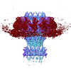 | |||||||||||||||
|---|---|---|---|---|---|---|---|---|---|---|---|---|---|---|---|---|
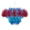
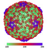
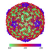
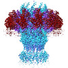
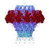
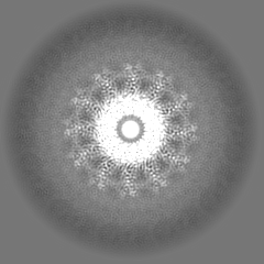
| Title | SaPI1 portal-capsid interface in mature capsids with DNA | |||||||||||||||
 Map data Map data | ||||||||||||||||
 Sample Sample |
| |||||||||||||||
 Keywords Keywords |  capsid / phage / sapi / portal / STRUCTURAL PROTEIN capsid / phage / sapi / portal / STRUCTURAL PROTEIN | |||||||||||||||
| Biological species |  Dubowvirus dv80alpha Dubowvirus dv80alpha | |||||||||||||||
| Method | single particle reconstruction / cryo EM / Resolution: 3.4 Å | |||||||||||||||
 Authors Authors | Kizziah JL / Mukherjee A / Dokland T | |||||||||||||||
| Funding support |  United States, 4 items United States, 4 items
| |||||||||||||||
 Citation Citation | Journal: J Mol Biol / Year: 2024 Title: Structure of the Portal Complex from Staphylococcus aureus Pathogenicity Island 1 Transducing Particles In Situ and In Isolation. Authors: Amarshi Mukherjee / James L Kizziah / N'Toia C Hawkins / Mohamed O Nasef / Laura K Parker / Terje Dokland / Abstract: Staphylococcus aureus is an important human pathogen, and the prevalence of antibiotic resistance is a major public health concern. The evolution of pathogenicity and resistance in S. aureus often ...Staphylococcus aureus is an important human pathogen, and the prevalence of antibiotic resistance is a major public health concern. The evolution of pathogenicity and resistance in S. aureus often involves acquisition of mobile genetic elements (MGEs). Bacteriophages play an especially important role, since transduction represents the main mechanism for horizontal gene transfer. S. aureus pathogenicity islands (SaPIs), including SaPI1, are MGEs that carry genes encoding virulence factors, and are mobilized at high frequency through interactions with specific "helper" bacteriophages, such as 80α, leading to packaging of the SaPI genomes into virions made from structural proteins supplied by the helper. Among these structural proteins is the portal protein, which forms a ring-like portal at a fivefold vertex of the capsid, through which the DNA is packaged during virion assembly and ejected upon infection of the host. We have used high-resolution cryo-electron microscopy to determine structures of the S. aureus bacteriophage 80α portal itself, produced by overexpression, and in situ in the empty and full SaPI1 virions, and show how the portal interacts with the capsid. These structures provide a basis for understanding portal and capsid assembly and the conformational changes that occur upon DNA packaging and ejection. | |||||||||||||||
| History |
|
- Structure visualization
| Supplemental images |
|---|

- Downloads & links
-EMDB archive
| Map data | emd_43147.map.gz | 49.4 MB |  EMDB map data format EMDB map data format | |
|---|---|---|---|---|
| Header (meta data) | emd-43147-v30.xmlemd-43147.xml | 21 KB 21 KB | Display Display | EMDB header |
| FSC (resolution estimation) | emd_43147_fsc.xml | 7.9 KB | Display | FSC data file |
| Images | 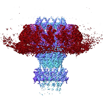 emd_43147.png emd_43147.png | 156.4 KB | ||
| Masks | emd_43147_msk_1.map | 52.7 MB | Mask map | |
| Filedesc metadata | emd-43147.cif.gz | 6 KB | ||
| Others | emd_43147_additional_1.map.gzemd_43147_additional_2.map.gzemd_43147_half_map_1.map.gzemd_43147_half_map_2.map.gz | 45.9 MB 44.8 MB 48.2 MB 48.2 MB | ||
| Archive directory |  http://ftp.pdbj.org/pub/emdb/structures/EMD-43147ftp://ftp.pdbj.org/pub/emdb/structures/EMD-43147 http://ftp.pdbj.org/pub/emdb/structures/EMD-43147ftp://ftp.pdbj.org/pub/emdb/structures/EMD-43147 | HTTPS FTP |
-Related structure data
| Related structure data |  8vdeMC  8v8bC 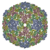 8vd4C 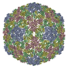 8vd5C  8vd8C  8vdcC C: citing same article ( M: atomic model generated by this map |
|---|
-Links
| EMDB pages | EMDB (EBI/PDBe) / EMDataResource |
|---|
-Map
| File | Download / File: emd_43147.map.gz / Format: CCP4 / Size: 52.7 MB / Type: IMAGE STORED AS FLOATING POINT NUMBER (4 BYTES) | ||||||||||||||||||||
|---|---|---|---|---|---|---|---|---|---|---|---|---|---|---|---|---|---|---|---|---|---|
| Voxel size | X=Y=Z: 1.33 Å | ||||||||||||||||||||
| Density |
| ||||||||||||||||||||
| Symmetry | Space group: 1 | ||||||||||||||||||||
| Details | EMDB XML:
|
-Supplemental data
-Mask #1
| File | emd_43147_msk_1.map | ||||||||||||
|---|---|---|---|---|---|---|---|---|---|---|---|---|---|
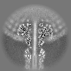
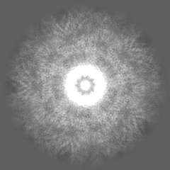
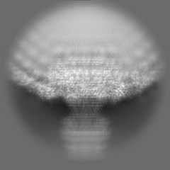
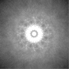
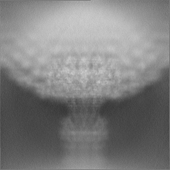
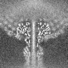
| Projections & Slices |
| ||||||||||||
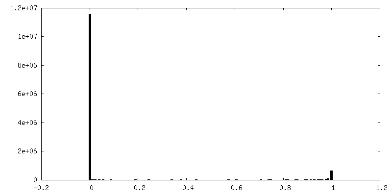
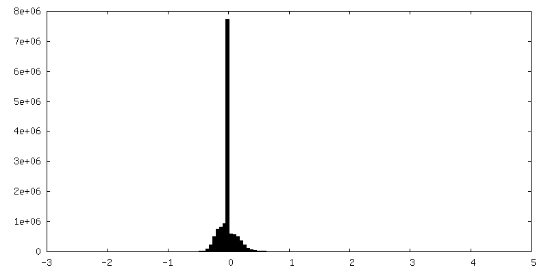
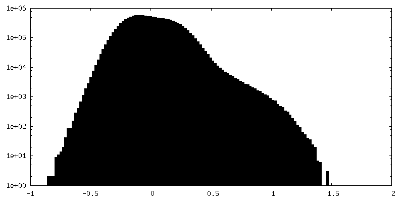
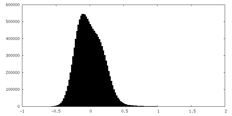
| Density Histograms |
 Z
Z Y
Y X
X


-Additional map: C12 symmetry averaged map resampled to main map...
| File | emd_43147_additional_1.map | ||||||||||||
|---|---|---|---|---|---|---|---|---|---|---|---|---|---|
| Annotation | C12 symmetry averaged map resampled to main map used to constrain portal protein models in ISOLDE | ||||||||||||
| Projections & Slices |
| ||||||||||||
| Density Histograms |


-Additional map: C5 symmetry averaged map resampled to main map...
| File | emd_43147_additional_2.map | ||||||||||||
|---|---|---|---|---|---|---|---|---|---|---|---|---|---|
| Annotation | C5 symmetry averaged map resampled to main map used to constrain capsid protein models in ISOLDE | ||||||||||||
| Projections & Slices |
| ||||||||||||
| Density Histograms |


-Half map: #2
| File | emd_43147_half_map_1.map | ||||||||||||
|---|---|---|---|---|---|---|---|---|---|---|---|---|---|
| Projections & Slices |
| ||||||||||||
| Density Histograms |


-Half map: #1
| File | emd_43147_half_map_2.map | ||||||||||||
|---|---|---|---|---|---|---|---|---|---|---|---|---|---|
| Projections & Slices |
| ||||||||||||
| Density Histograms |


- Sample components
-Entire : Dubowvirus dv80alpha
| Entire | Name: Dubowvirus dv80alpha |
|---|---|
| Components |
|
-Supramolecule #1: Dubowvirus dv80alpha
| Supramolecule | Name: Dubowvirus dv80alpha / type: virus / ID: 1 / Parent: 0 / Macromolecule list: all / NCBI-ID: 53369 / Sci species name: Dubowvirus dv80alpha / Virus type: SATELLITE / Virus isolate: SPECIES / Virus enveloped: No / Virus empty: No |
|---|
-Macromolecule #1: Major capsid protein
| Macromolecule | Name: Major capsid protein / type: protein_or_peptide / ID: 1 / Number of copies: 15 / Enantiomer: LEVO |
|---|---|
| Source (natural) | Organism: Dubowvirus dv80alpha |
| Molecular weight | Theoretical: 36.846883 KDa |
| Recombinant expression | Organism:  Escherichia coli (E. coli) Escherichia coli (E. coli) |
| Sequence | String: MEQTQKLKLN LQHFASNNVK PQVFNPDNVM MHEKKDGTLM NEFTTPILQE VMENSKIMQL GKYEPMEGTE KKFTFWADKP GAYWVGEGQ KIETSKATWV NATMRAFKLG VILPVTKEFL NYTYSQFFEE MKPMIAEAFY KKFDEAGILN QGNNPFGKSI A QSIEKTNK ...String: MEQTQKLKLN LQHFASNNVK PQVFNPDNVM MHEKKDGTLM NEFTTPILQE VMENSKIMQL GKYEPMEGTE KKFTFWADKP GAYWVGEGQ KIETSKATWV NATMRAFKLG VILPVTKEFL NYTYSQFFEE MKPMIAEAFY KKFDEAGILN QGNNPFGKSI A QSIEKTNK VIKGDFTQDN IIDLEALLED DELEANAFIS KTQNRSLLRK IVDPETKERI YDRNSDSLDG LPVVNLKSSN LK RGELITG DFDKLIYGIP QLIEYKIDET AQLSTVKNED GTPVNLFEQD MVALRATMHV ALHIADDKAF AKLVPADKRT DSV PGEV |
-Macromolecule #2: Portal protein
| Macromolecule | Name: Portal protein / type: protein_or_peptide / ID: 2 / Number of copies: 12 / Enantiomer: LEVO |
|---|---|
| Source (natural) | Organism: Dubowvirus dv80alpha |
| Molecular weight | Theoretical: 59.55182 KDa |
| Recombinant expression | Organism: Escherichia coli (E. coli) |
| Sequence | String: MLKVNEFETD TDLRGNINYL FNDEANVVYT YDGTESDLLQ NVNEVSKYIE HHMDYQRPRL KVLSDYYEGK TKNLVELTRR KEEYMADNR VAHDYASYIS DFINGYFLGN PIQYQDDDKD VLEAIEAFND LNDVESHNRS LGLDLSIYGK AYELMIRNQD D ETRLYKSD ...String: MLKVNEFETD TDLRGNINYL FNDEANVVYT YDGTESDLLQ NVNEVSKYIE HHMDYQRPRL KVLSDYYEGK TKNLVELTRR KEEYMADNR VAHDYASYIS DFINGYFLGN PIQYQDDDKD VLEAIEAFND LNDVESHNRS LGLDLSIYGK AYELMIRNQD D ETRLYKSD AMSTFIIYDN TVERNSIAGV RYLRTKPIDK TDEDEVFTVD LFTSHGVYRY LTNRTNGLKL TPRENSFESH SF ERMPITE FSNNERRKGD YEKVITLIDL YDNAESDTAN YMSDLNDAML LIKGNLNLDP VEVRKQKEAN VLFLEPTVYV DAE GRETEG SVDGGYIYKQ YDVQGTEAYK DRLNSDIHMF TNTPNMKDDN FSGTQSGEAM KYKLFGLEQR TKTKEGLFTK GLRR RAKLL ETILKNTRSI DANKDFNTVR YVYNRNLPKS LIEELKAYID SGGKISQTTL MSLFSFFQDP ELEVKKIEED EKESI KKAQ KGIYKDPRDI NDDEQDDDTK DTVDKKE |
-Experimental details
-Structure determination
| Method | cryo EM |
|---|---|
 Processing Processing | single particle reconstruction |
| Aggregation state | particle |
-Sample preparation
| Buffer | pH: 7.8 |
|---|---|
| Vitrification | Cryogen name: ETHANE |
- Electron microscopy
| Microscope | FEI TITAN KRIOS |
|---|---|
| Electron beam | Acceleration voltage: 300 kV / Electron source: FIELD EMISSION GUN |
| Electron optics | Illumination mode: FLOOD BEAM / Imaging mode: BRIGHT FIELDBright-field microscopy / Nominal defocus max: 2.0 µm / Nominal defocus min: 0.7000000000000001 µm |
| Image recording | Film or detector model: GATAN K3 BIOQUANTUM (6k x 4k) / Average electron dose: 35.26 e/Å2 |
| Experimental equipment |  Model: Titan Krios / Image courtesy: FEI Company |
-Image processing
| Startup model | Type of model: INSILICO MODEL |
|---|---|
| Initial angle assignment | Type: RANDOM ASSIGNMENT |
| Final angle assignment | Type: MAXIMUM LIKELIHOOD |
| Final reconstruction | Resolution.type: BY AUTHOR / Resolution: 3.4 Å / Resolution method: FSC 0.143 CUT-OFF / Number images used: 59457 |
| FSC plot (resolution estimation) |  |
