 Movie
Movie Controller


+ Open data

- Basic information
| Entry | Database: EMDB / ID: EMD-4263 | |||||||||
|---|---|---|---|---|---|---|---|---|---|---|
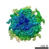
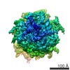
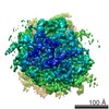
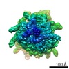
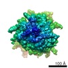
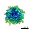
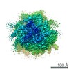
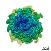
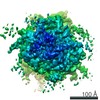
| Title | High-resolution cryo-EM structure of the human 80S ribosome | |||||||||
 Map data Map data | Human 80S ribosomes 3D reconstruction before focused refinement (Corresponding high-resolution obtained by focused refinement is EMD-3883) | |||||||||
 Sample Sample |
| |||||||||
| Biological species |  Homo sapiens (human) Homo sapiens (human) | |||||||||
| Method | single particle reconstruction / cryo EM / Resolution: 2.9 Å | |||||||||
 Authors Authors | Natchiar SK / Myasnikov AG | |||||||||
 Citation Citation | Journal: Nature / Year: 2017 Title: Visualization of chemical modifications in the human 80S ribosome structure. Authors: S Kundhavai Natchiar / Alexander G Myasnikov / Hanna Kratzat / Isabelle Hazemann / Bruno P Klaholz /  Abstract: Chemical modifications of human ribosomal RNA (rRNA) are introduced during biogenesis and have been implicated in the dysregulation of protein synthesis, as is found in cancer and other diseases. ...Chemical modifications of human ribosomal RNA (rRNA) are introduced during biogenesis and have been implicated in the dysregulation of protein synthesis, as is found in cancer and other diseases. However, their role in this phenomenon is unknown. Here we visualize more than 130 individual rRNA modifications in the three-dimensional structure of the human ribosome, explaining their structural and functional roles. In addition to a small number of universally conserved sites, we identify many eukaryote- or human-specific modifications and unique sites that form an extended shell in comparison to bacterial ribosomes, and which stabilize the RNA. Several of the modifications are associated with the binding sites of three ribosome-targeting antibiotics, or are associated with degenerate states in cancer, such as keto alkylations on nucleotide bases reminiscent of specialized ribosomes. This high-resolution structure of the human 80S ribosome paves the way towards understanding the role of epigenetic rRNA modifications in human diseases and suggests new possibilities for designing selective inhibitors and therapeutic drugs. | |||||||||
| History |
|
- Structure visualization
| Movie |
 Movie viewer Movie viewer |
|---|---|
| Structure viewer | EM map: SurfViewMolmilJmol/JSmol |
| Supplemental images |

- Downloads & links
-EMDB archive
| Map data | emd_4263.map.gz | 809.8 MB | EMDB map data format | |
|---|---|---|---|---|
| Header (meta data) | emd-4263-v30.xmlemd-4263.xml | 23.8 KB 23.8 KB | Display Display | EMDB header |
| Images | 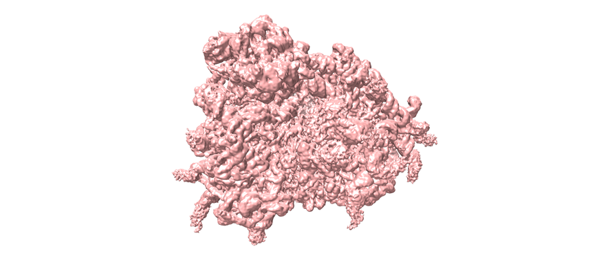 emd_4263.png emd_4263.png | 191.3 KB | ||
| Masks | emd_4263_msk_1.map | 1000 MB | Mask map | |
| Others | emd_4263_half_map_1.map.gzemd_4263_half_map_2.map.gz | 809.8 MB 810.2 MB | ||
| Archive directory |  http://ftp.pdbj.org/pub/emdb/structures/EMD-4263ftp://ftp.pdbj.org/pub/emdb/structures/EMD-4263 http://ftp.pdbj.org/pub/emdb/structures/EMD-4263ftp://ftp.pdbj.org/pub/emdb/structures/EMD-4263 | HTTPS FTP |
-Validation report
| Summary document | emd_4263_validation.pdf.gz | 435 KB | Display | EMDB validaton report |
|---|---|---|---|---|
| Full document | emd_4263_full_validation.pdf.gz | 434.1 KB | Display | |
| Data in XML | emd_4263_validation.xml.gz | 18.8 KB | Display | |
| Arichive directory | https://ftp.pdbj.org/pub/emdb/validation_reports/EMD-4263ftp://ftp.pdbj.org/pub/emdb/validation_reports/EMD-4263 | HTTPS FTP |
-Related structure data


-Links
| EMDB pages | EMDB (EBI/PDBe) / EMDataResource |
|---|---|
| Related items in Molecule of the Month |

-Map
| File | Download / File: emd_4263.map.gz / Format: CCP4 / Size: 1000 MB / Type: IMAGE STORED AS FLOATING POINT NUMBER (4 BYTES) | ||||||||||||||||||||||||||||||||||||||||||||||||||||||||||||||||||||
|---|---|---|---|---|---|---|---|---|---|---|---|---|---|---|---|---|---|---|---|---|---|---|---|---|---|---|---|---|---|---|---|---|---|---|---|---|---|---|---|---|---|---|---|---|---|---|---|---|---|---|---|---|---|---|---|---|---|---|---|---|---|---|---|---|---|---|---|---|---|
| Annotation | Human 80S ribosomes 3D reconstruction before focused refinement (Corresponding high-resolution obtained by focused refinement is EMD-3883) | ||||||||||||||||||||||||||||||||||||||||||||||||||||||||||||||||||||
| Voxel size | X=Y=Z: 0.85 Å | ||||||||||||||||||||||||||||||||||||||||||||||||||||||||||||||||||||
| Density |
| ||||||||||||||||||||||||||||||||||||||||||||||||||||||||||||||||||||
| Symmetry | Space group: 1 | ||||||||||||||||||||||||||||||||||||||||||||||||||||||||||||||||||||
| Details | EMDB XML:
CCP4 map header:
| ||||||||||||||||||||||||||||||||||||||||||||||||||||||||||||||||||||
-Supplemental data
-Mask #1
| File | emd_4263_msk_1.map | ||||||||||||
|---|---|---|---|---|---|---|---|---|---|---|---|---|---|
| Projections & Slices |
| ||||||||||||
| Density Histograms |
 Z
Z Y
Y X
X


-Half map: Human 80S ribosomes half map before focused refinement...
| File | emd_4263_half_map_1.map | ||||||||||||
|---|---|---|---|---|---|---|---|---|---|---|---|---|---|
| Annotation | Human 80S ribosomes half map before focused refinement (Corresponding high-resolution obtained by focused refinement is EMD-3883) | ||||||||||||
| Projections & Slices |
| ||||||||||||
| Density Histograms |


-Half map: Human 80S ribosomes half map before focused refinement...
| File | emd_4263_half_map_2.map | ||||||||||||
|---|---|---|---|---|---|---|---|---|---|---|---|---|---|
| Annotation | Human 80S ribosomes half map before focused refinement (Corresponding high-resolution obtained by focused refinement is EMD-3883) | ||||||||||||
| Projections & Slices |
| ||||||||||||
| Density Histograms |


- Sample components
-Entire : 80S ribosome with ligand HHT and HYG
| Entire | Name: 80S ribosome with ligand HHT and HYG |
|---|---|
| Components |
|
-Supramolecule #1: 80S ribosome with ligand HHT and HYG
| Supramolecule | Name: 80S ribosome with ligand HHT and HYG / type: complex / ID: 1 / Parent: 0 / Macromolecule list: #1-#81 / Details: Human 80S ribosome with modified nucleotides |
|---|---|
| Source (natural) | Organism: Homo sapiens (human) |
| Molecular weight | Experimental: 4.3 MDa |
-Experimental details
-Structure determination
| Method | cryo EM |
|---|---|
 Processing Processing | single particle reconstruction |
| Aggregation state | particle |
-Sample preparation
| Concentration | 0.5 mg/mL |
|---|---|
| Buffer | pH: 7.5 / Details: 100mM KCl, 5mM MgAc2, 20mM Hepes, 10mM NH4Cl |
| Vitrification | Cryogen name: ETHANE / Chamber humidity: 100 % / Chamber temperature: 283 K / Instrument: FEI VITROBOT MARK IV |
- Electron microscopy
| Microscope | FEI TITAN KRIOS |
|---|---|
| Image recording | Film or detector model: FEI FALCON II (4k x 4k) / Average electron dose: 3.5 e/Å2 |
| Electron beam | Acceleration voltage: 300 kV / Electron source:  FIELD EMISSION GUN FIELD EMISSION GUN |
| Electron optics | Illumination mode: SPOT SCAN / Imaging mode: BRIGHT FIELD |
| Sample stage | Specimen holder model: FEI TITAN KRIOS AUTOGRID HOLDER |
| Experimental equipment |  Model: Titan Krios / Image courtesy: FEI Company |
-Image processing
| Final reconstruction | Resolution.type: BY AUTHOR / Resolution: 2.9 Å / Resolution method: FSC 0.143 CUT-OFF / Number images used: 138234 |
|---|---|
| Initial angle assignment | Type: NOT APPLICABLE |
| Final angle assignment | Type: NOT APPLICABLE |
-Atomic model buiding 1
| Refinement | Protocol: OTHER |
|---|
