 Movie
Movie Controller

[English] 日本語
 Yorodumi
- EMDB-41761: Cryo-EM structure of Vibrio cholerae FtsE/FtsX/EnvC complex, shortened -
+ Open data

- Basic information
| Entry |  | |||||||||
|---|---|---|---|---|---|---|---|---|---|---|
| Title | Cryo-EM structure of Vibrio cholerae FtsE/FtsX/EnvC complex, shortened | |||||||||
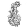
 Map data Map data | full map | |||||||||
 Sample Sample |
| |||||||||
 Keywords Keywords |  membrane protein / enzyme / TRANSPORT PROTEIN membrane protein / enzyme / TRANSPORT PROTEIN | |||||||||
| Function / homology |  Function and homology informationcell cycle / cell division / ATP hydrolysis activity / ATP binding / plasma membrane Function and homology informationcell cycle / cell division / ATP hydrolysis activity / ATP binding / plasma membraneSimilarity search - Function | |||||||||
| Biological species |   Vibrio cholerae (bacteria) Vibrio cholerae (bacteria) | |||||||||
| Method | single particle reconstruction / cryo EM / Resolution: 3.55 Å | |||||||||
 Authors Authors | Hao A / Lee S-Y | |||||||||
| Funding support |  United States, 1 items United States, 1 items
| |||||||||
 Citation Citation | Journal: Structure / Year: 2024 Title: Structural insights into the FtsEX-EnvC complex regulation on septal peptidoglycan hydrolysis in Vibrio cholerae. Authors: Aili Hao / Yang Suo / Seok-Yong Lee / Abstract: During bacterial cell division, hydrolysis of septal peptidoglycan (sPG) is crucial for cell separation. This sPG hydrolysis is performed by the enzyme amidases whose activity is regulated by the ...During bacterial cell division, hydrolysis of septal peptidoglycan (sPG) is crucial for cell separation. This sPG hydrolysis is performed by the enzyme amidases whose activity is regulated by the integral membrane protein complex FtsEX-EnvC. FtsEX is an ATP-binding cassette transporter, and EnvC is a long coiled-coil protein that interacts with and activates the amidases. The molecular mechanism by which the FtsEX-EnvC complex activates amidases remains largely unclear. We present the cryo-electron microscopy structure of the FtsEX-EnvC complex from the pathogenic bacteria V. cholerae (FtsEX-EnvC). FtsEX-EnvC in the presence of ADP adopts a distinct conformation where EnvC is "horizontally extended" rather than "vertically extended". Subsequent structural studies suggest that EnvC can swing between these conformations in space in a nucleotide-dependent manner. Our structural analysis and functional studies suggest that FtsEX-EnvC employs spatial control of EnvC for amidase activation, providing mechanistic insights into the FtsEX-EnvC regulation on septal peptidoglycan hydrolysis. | |||||||||
| History |
|
- Structure visualization
| Supplemental images |
|---|

- Downloads & links
-EMDB archive
| Map data | emd_41761.map.gz | 179.4 MB | EMDB map data format | |
|---|---|---|---|---|
| Header (meta data) | emd-41761-v30.xmlemd-41761.xml | 21 KB 21 KB | Display Display | EMDB header |
| Images |  emd_41761.png emd_41761.png | 80.1 KB | ||
| Masks | emd_41761_msk_1.map | 347.6 MB | Mask map | |
| Filedesc metadata | emd-41761.cif.gz | 6.9 KB | ||
| Others | emd_41761_half_map_1.map.gzemd_41761_half_map_2.map.gz | 323 MB 323.1 MB | ||
| Archive directory |  http://ftp.pdbj.org/pub/emdb/structures/EMD-41761ftp://ftp.pdbj.org/pub/emdb/structures/EMD-41761 http://ftp.pdbj.org/pub/emdb/structures/EMD-41761ftp://ftp.pdbj.org/pub/emdb/structures/EMD-41761 | HTTPS FTP |
-Related structure data
| Related structure data |  8tzkMC 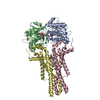 8tzjC  8tzlC C: citing same article ( M: atomic model generated by this map |
|---|---|
| Similar structure data |


-Links
| EMDB pages | EMDB (EBI/PDBe) / EMDataResource |
|---|---|
| Related items in Molecule of the Month |


-Map
| File | Download / File: emd_41761.map.gz / Format: CCP4 / Size: 347.6 MB / Type: IMAGE STORED AS FLOATING POINT NUMBER (4 BYTES) | ||||||||||||||||||||
|---|---|---|---|---|---|---|---|---|---|---|---|---|---|---|---|---|---|---|---|---|---|
| Annotation | full map | ||||||||||||||||||||
| Voxel size | X=Y=Z: 1.08 Å | ||||||||||||||||||||
| Density |
| ||||||||||||||||||||
| Symmetry | Space group: 1 | ||||||||||||||||||||
| Details | EMDB XML:
|
-Supplemental data
-Mask #1
| File | emd_41761_msk_1.map | ||||||||||||
|---|---|---|---|---|---|---|---|---|---|---|---|---|---|
| Projections & Slices |
| ||||||||||||
| Density Histograms |
 Z
Z Y
Y X
X


-Half map: half map 2
| File | emd_41761_half_map_1.map | ||||||||||||
|---|---|---|---|---|---|---|---|---|---|---|---|---|---|
| Annotation | half map 2 | ||||||||||||
| Projections & Slices |
| ||||||||||||
| Density Histograms |


-Half map: half map 1
| File | emd_41761_half_map_2.map | ||||||||||||
|---|---|---|---|---|---|---|---|---|---|---|---|---|---|
| Annotation | half map 1 | ||||||||||||
| Projections & Slices |
| ||||||||||||
| Density Histograms |


- Sample components
-Entire : FtsEX
| Entire | Name: FtsEX |
|---|---|
| Components |
|
-Supramolecule #1: FtsEX
| Supramolecule | Name: FtsEX / type: complex / ID: 1 / Parent: 0 / Macromolecule list: #1-#3 |
|---|---|
| Source (natural) | Organism: Vibrio cholerae (bacteria) |
| Molecular weight | Theoretical: 200 KDa |
-Macromolecule #1: Cell division ATP-binding protein FtsE
| Macromolecule | Name: Cell division ATP-binding protein FtsE / type: protein_or_peptide / ID: 1 / Number of copies: 2 / Enantiomer: LEVO |
|---|---|
| Source (natural) | Organism: Vibrio cholerae (bacteria) |
| Molecular weight | Theoretical: 25.882668 KDa |
| Recombinant expression | Organism: Escherichia coli (E. coli) |
| Sequence | String: PALSGGDGVI RFQQVSKAYR GGRQALQKVD FHLRRGEMAF LGGHSGAGKS TLLKLICAIE RPTDGKISFN GHDITRIPNK DIPFLRRNI GIVFQDHRLL MDRSIYDNVA LPMRIESISE NEIKRRVSAA LDKTGLLDKA RCLPSQLSGG EQQRVGIARA V VNRPTLLL ...String: PALSGGDGVI RFQQVSKAYR GGRQALQKVD FHLRRGEMAF LGGHSGAGKS TLLKLICAIE RPTDGKISFN GHDITRIPNK DIPFLRRNI GIVFQDHRLL MDRSIYDNVA LPMRIESISE NEIKRRVSAA LDKTGLLDKA RCLPSQLSGG EQQRVGIARA V VNRPTLLL ADEPTGNLDP ELSSRVLRLF EEFNRAGVTI LLATHDIHLV NSRPQYRHLE LNQGFLSEVA DYGR UniProtKB: Cell division ATP-binding protein FtsE |
-Macromolecule #2: Cell division protein FtsX
| Macromolecule | Name: Cell division protein FtsX / type: protein_or_peptide / ID: 2 / Number of copies: 2 / Enantiomer: LEVO |
|---|---|
| Source (natural) | Organism: Vibrio cholerae (bacteria) |
| Molecular weight | Theoretical: 36.516773 KDa |
| Recombinant expression | Organism: Escherichia coli (E. coli) |
| Sequence | String: MAVKPGNQKI SKTTKSTKSK PRDVKRAKTD SFLAIHFKQA KASFAALWRR PLGNILTLAV ISMALALPAS LYLLSKNIAS VAERVAEPS QLSVYLHIDT PEPRIIVLKD DLERRDEIAK VKYISPQQGL DDLSQYAGFE QAISLLDNAT LPAVLVVTPK V DSREQIQT ...String: MAVKPGNQKI SKTTKSTKSK PRDVKRAKTD SFLAIHFKQA KASFAALWRR PLGNILTLAV ISMALALPAS LYLLSKNIAS VAERVAEPS QLSVYLHIDT PEPRIIVLKD DLERRDEIAK VKYISPQQGL DDLSQYAGFE QAISLLDNAT LPAVLVVTPK V DSREQIQT LAKALQAEEG VTDVRMDEDW FARLDAIRHL ATIVVISLSS LMLMSVFLIV GNTLRFNVQA NKEEIQTMKL IG ATDAYIL RPYLYSGMWF GLLGAVAAWL LTALMTILLN GAVEALAQLY DSRFRLIGLG WDESLLLLML GVFLGCVAAK VSA KRHLKE IEPV UniProtKB: Cell division protein FtsX |
-Macromolecule #3: Peptidase M23
| Macromolecule | Name: Peptidase M23 / type: protein_or_peptide / ID: 3 / Number of copies: 1 / Enantiomer: LEVO |
|---|---|
| Source (natural) | Organism: Vibrio cholerae (bacteria) |
| Molecular weight | Theoretical: 43.410895 KDa |
| Recombinant expression | Organism: Escherichia coli (E. coli) |
| Sequence | String: MTATDPHAIF SDFLGKTLTH RLLACLLFMV SPSLFAATQQ ELTGVKSEIS RQQQSLAEQQ KSLDQLQQAL KQQELGINSI ENQITKTKN DLENANRNIA QLNSNIQALE TQKQQQADKL ERLLQTYYLT KRSLTNGQFF HRSADEDRIS QYYQHLAKSR A QAIEALEK ...String: MTATDPHAIF SDFLGKTLTH RLLACLLFMV SPSLFAATQQ ELTGVKSEIS RQQQSLAEQQ KSLDQLQQAL KQQELGINSI ENQITKTKN DLENANRNIA QLNSNIQALE TQKQQQADKL ERLLQTYYLT KRSLTNGQFF HRSADEDRIS QYYQHLAKSR A QAIEALEK TQTELNSNQK QRQTEREQIE KLLAEQTQQR DKLAKTQSER KQTVKKIESS ISGNKTYLAE LQRNETRLKA EI AKAAKRN AVLMNGIASQ RGKLPWPLKG RVLHNFGERQ TGQIDWKGLV IDANYGQEVK AVYPGTIVFA EYLRGYGLVV LLD HGKGDM TLYGFNQTLL KKEGDKVTTG ETIALAGDTG GQSRPALYFE IRRNSRAENP SQWLQR UniProtKB: Peptidase M23 |
-Macromolecule #4: MAGNESIUM ION
| Macromolecule | Name: MAGNESIUM ION / type: ligand / ID: 4 / Number of copies: 2 / Formula: MG |
|---|---|
| Molecular weight | Theoretical: 24.305 Da |
-Macromolecule #5: ADENOSINE-5'-DIPHOSPHATE
| Macromolecule | Name: ADENOSINE-5'-DIPHOSPHATE / type: ligand / ID: 5 / Number of copies: 2 / Formula: ADP |
|---|---|
| Molecular weight | Theoretical: 427.201 Da |
| Chemical component information |  ChemComp-ADP: |
-Experimental details
-Structure determination
| Method | cryo EM |
|---|---|
 Processing Processing | single particle reconstruction |
| Aggregation state | particle |
-Sample preparation
| Concentration | 8 mg/mL | |||||||||
|---|---|---|---|---|---|---|---|---|---|---|
| Buffer | pH: 8 Component:
| |||||||||
| Vitrification | Cryogen name: ETHANE / Chamber humidity: 95 % / Chamber temperature: 277 K / Instrument: LEICA EM GP |
- Electron microscopy
| Microscope | FEI TITAN KRIOS |
|---|---|
| Electron beam | Acceleration voltage: 300 kV / Electron source: FIELD EMISSION GUN |
| Electron optics | C2 aperture diameter: 100.0 µm / Illumination mode: FLOOD BEAM / Imaging mode: BRIGHT FIELDBright-field microscopy / Cs: 2.7 mm / Nominal defocus max: 2.0 µm / Nominal defocus min: 1.0 µm / Nominal magnification: 81000 |
| Specialist optics | Energy filter - Name: GIF Bioquantum / Energy filter - Slit width: 20 eV |
| Sample stage | Specimen holder model: FEI TITAN KRIOS AUTOGRID HOLDER / Cooling holder cryogen: NITROGEN |
| Image recording | Film or detector model: GATAN K3 BIOQUANTUM (6k x 4k) / Digitization - Dimensions - Width: 5760 pixel / Digitization - Dimensions - Height: 4092 pixel / Number grids imaged: 1 / Average exposure time: 2.4 sec. / Average electron dose: 60.0 e/Å2 |
| Experimental equipment |  Model: Titan Krios / Image courtesy: FEI Company |
-Image processing
| Particle selection | Number selected: 2434011 |
|---|---|
| Startup model | Type of model: OTHER |
| Initial angle assignment | Type: OTHER / Software - Name: cryoSPARC (ver. 3.3) |
| Final 3D classification | Number classes: 4 / Software - Name: cryoSPARC (ver. 3.3) |
| Final angle assignment | Type: OTHER / Software - Name: cryoSPARC (ver. 3.3) |
| Final reconstruction | Number classes used: 1 / Algorithm: FOURIER SPACE / Resolution.type: BY AUTHOR / Resolution: 3.55 Å / Resolution method: FSC 0.143 CUT-OFF / Software - Name: cryoSPARC (ver. 3.3) / Number images used: 179239 |
-Atomic model buiding 1
| Initial model | Chain - Source name: AlphaFold / Chain - Initial model type: in silico model |
|---|---|
| Refinement | Space: REAL / Protocol: FLEXIBLE FIT |
| Output model | PDB-8tzk: |
