National Institutes of Health/National Institute Of Allergy and Infectious Diseases (NIH/NIAID)
U54-AI170856
United States
Citation
Journal: J Mol Biol / Year: 2024 Title: Molecular Determinants of PQBP1 Binding to the HIV-1 Capsid Lattice. Authors: Juliana Piacentini / Dale S Allen / Barbie K Ganser-Pornillos / Sumit K Chanda / Sunnie M Yoh / Owen Pornillos / Abstract: Human immunodeficiency virus type 1 (HIV-1) stimulates innate immune responses upon infection, including cyclic GMP-AMP synthase (cGAS) signaling that results in type I interferon production. HIV-1- ...Human immunodeficiency virus type 1 (HIV-1) stimulates innate immune responses upon infection, including cyclic GMP-AMP synthase (cGAS) signaling that results in type I interferon production. HIV-1-induced activation of cGAS requires the host cell factor polyglutamine binding protein 1 (PQBP1), an intrinsically disordered protein that bridges capsid recognition and cGAS recruitment. However, the molecular details of PQBP1 interactions with the HIV-1 capsid and their functional implications remain poorly understood. Here, we show that PQBP1 binds to HIV-1 capsids through charge complementing contacts between acidic residues in the N-terminal region of PQBP1 and an arginine ring in the central channel of the HIV-1 CA hexamer that makes up the viral capsid. These studies reveal the molecular details of PQBP1's primary interaction with the HIV-1 capsid and suggest that additional elements are likely to contribute to stable capsid binding.
In the structure databanks used in Yorodumi, some data are registered as the other names, "COVID-19 virus" and "2019-nCoV". Here are the details of the virus and the list of structure data.
Jan 31, 2019. EMDB accession codes are about to change! (news from PDBe EMDB page)
EMDB accession codes are about to change! (news from PDBe EMDB page)
The allocation of 4 digits for EMDB accession codes will soon come to an end. Whilst these codes will remain in use, new EMDB accession codes will include an additional digit and will expand incrementally as the available range of codes is exhausted. The current 4-digit format prefixed with “EMD-” (i.e. EMD-XXXX) will advance to a 5-digit format (i.e. EMD-XXXXX), and so on. It is currently estimated that the 4-digit codes will be depleted around Spring 2019, at which point the 5-digit format will come into force.
The EM Navigator/Yorodumi systems omit the EMD- prefix.
Related info.:Q: What is EMD? / ID/Accession-code notation in Yorodumi/EM Navigator
Yorodumi is a browser for structure data from EMDB, PDB, SASBDB, etc.
This page is also the successor to EM Navigator detail page, and also detail information page/front-end page for Omokage search.
The word "yorodu" (or yorozu) is an old Japanese word meaning "ten thousand". "mi" (miru) is to see.
Related info.:EMDB / PDB / SASBDB / Comparison of 3 databanks / Yorodumi Search / Aug 31, 2016. New EM Navigator & Yorodumi / Yorodumi Papers / Jmol/JSmol / Function and homology information / Changes in new EM Navigator and Yorodumi
 Movie
Movie Controller


 Open data
 Basic information
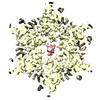
 Map data
 Sample
 Keywords
 capsid / innate immune sensor /
  Function and homology information

 Authors
 United States, 1 items
  Citation
 Structure visualization
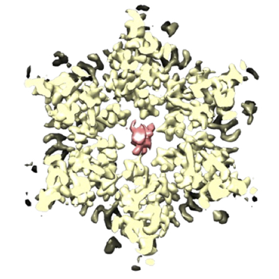
 Downloads & links
 emd_41711.png
 http://ftp.pdbj.org/pub/emdb/structures/EMD-41711
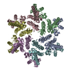


 Z
Z Y
Y X
X


 Sample components
 Processing
 Electron microscopy


