 Movie
Movie Controller


+ Open data

- Basic information
| Entry | Database: EMDB / ID: EMD-4158 | |||||||||
|---|---|---|---|---|---|---|---|---|---|---|
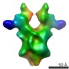
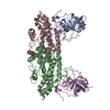
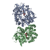
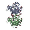
| Title | PilMN complex from Thermus thermophilus | |||||||||
 Map data Map data | Thermus PilMN complex | |||||||||
 Sample Sample |
| |||||||||
| Biological species |    Thermus thermophilus HB8 (bacteria) Thermus thermophilus HB8 (bacteria) | |||||||||
| Method | single particle reconstruction / cryo EM / Resolution: 27.0 Å | |||||||||
 Authors Authors | Derrick JP / Collins RF | |||||||||
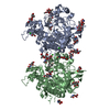
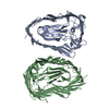
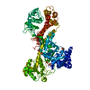
 Citation Citation | Journal: Proc Natl Acad Sci U S A / Year: 2013 Title: Structure and assembly of an inner membrane platform for initiation of type IV pilus biogenesis. Authors: Vijaykumar Karuppiah / Richard F Collins / Angela Thistlethwaite / Ya Gao / Jeremy P Derrick /  Abstract: Type IV pili are long fibers that are assembled by polymerization of a major pilin protein in the periplasm of a wide range of bacteria and archaea. They play crucial roles in pathogenesis, DNA ...Type IV pili are long fibers that are assembled by polymerization of a major pilin protein in the periplasm of a wide range of bacteria and archaea. They play crucial roles in pathogenesis, DNA transformation, and motility, and are capable of rapid retraction, generating powerful motor forces. PilN and PilO are integral inner membrane proteins that are essential for type IV pilus formation. Here, we show that PilN and PilO from Thermus thermophilus can be isolated as a complex with PilM, a cytoplasmic protein with structural similarities to the cytoskeletal protein MreB. The crystal structure of the periplasmic portion of PilN forms a homodimer with an extensive, conserved interaction interface. We conducted serial 3D reconstructions by electron microscopy of PilMN, PilMNO, and PilMNO bound to the major pilin protein PilA4, to chart the assembly of the inner membrane pilus biogenesis platform. PilN drives the dimerization of the PilMN complex with a stoichiometry of 2:2; binding of two PilO monomers then causes the PilN periplasmic domains to dissociate. Finally, two PilA4 monomers bind to the periplasmic domains of PilN and PilO, to generate a T-shaped complex that is primed for addition of the pilin to the nascent pilus fiber. Docking of structures for PilM, PilN, PilO, and PilA4 into the electron density maps of the transmembrane complexes was used to generate a sequence of molecular structures that chart the initial events in type IV pilus formation, and provide structural information on the early events in this important secretion process. | |||||||||
| History |
|
- Structure visualization
| Movie |
 Movie viewer Movie viewer |
|---|---|
| Structure viewer | EM map: SurfViewMolmilJmol/JSmol |
| Supplemental images |

- Downloads & links
-EMDB archive
| Map data | emd_4158.map.gz | 114.7 KB | EMDB map data format | |
|---|---|---|---|---|
| Header (meta data) | emd-4158-v30.xmlemd-4158.xml | 10 KB 10 KB | Display Display | EMDB header |
| Images |  emd_4158.png emd_4158.png | 31.7 KB | ||
| Archive directory |  http://ftp.pdbj.org/pub/emdb/structures/EMD-4158ftp://ftp.pdbj.org/pub/emdb/structures/EMD-4158 http://ftp.pdbj.org/pub/emdb/structures/EMD-4158ftp://ftp.pdbj.org/pub/emdb/structures/EMD-4158 | HTTPS FTP |
-Related structure data


-Links
| EMDB pages | EMDB (EBI/PDBe) / EMDataResource |
|---|
-Map
| File | Download / File: emd_4158.map.gz / Format: CCP4 / Size: 1 MB / Type: IMAGE STORED AS FLOATING POINT NUMBER (4 BYTES) | ||||||||||||||||||||||||||||||||||||||||||||||||||||||||||||
|---|---|---|---|---|---|---|---|---|---|---|---|---|---|---|---|---|---|---|---|---|---|---|---|---|---|---|---|---|---|---|---|---|---|---|---|---|---|---|---|---|---|---|---|---|---|---|---|---|---|---|---|---|---|---|---|---|---|---|---|---|---|
| Annotation | Thermus PilMN complex | ||||||||||||||||||||||||||||||||||||||||||||||||||||||||||||
| Voxel size | X=Y=Z: 3.49 Å | ||||||||||||||||||||||||||||||||||||||||||||||||||||||||||||
| Density |
| ||||||||||||||||||||||||||||||||||||||||||||||||||||||||||||
| Symmetry | Space group: 1 | ||||||||||||||||||||||||||||||||||||||||||||||||||||||||||||
| Details | EMDB XML:
CCP4 map header:
| ||||||||||||||||||||||||||||||||||||||||||||||||||||||||||||
-Supplemental data
- Sample components
-Entire : PilMN complex
| Entire | Name: PilMN complex |
|---|---|
| Components |
|
-Supramolecule #1: PilMN complex
| Supramolecule | Name: PilMN complex / type: complex / ID: 1 / Parent: 0 / Macromolecule list: all |
|---|---|
| Source (natural) | Organism: Thermus thermophilus HB8 (bacteria) |
| Recombinant expression | Organism: Escherichia coli (E. coli) / Recombinant plasmid: pET28a |
-Macromolecule #1: PilN
| Macromolecule | Name: PilN / type: protein_or_peptide / ID: 1 / Enantiomer: LEVO |
|---|---|
| Source (natural) | Organism: Thermus thermophilus HB8 (bacteria) |
| Recombinant expression | Organism: Escherichia coli (E. coli) |
| Sequence | String: MIRLNLLPKN LRRRVEPGWW RLLALLFALV VLGVLGLVHY TAYTELSLAK AERDQLQAEV EALRPFIAE LGRLQEERKA LEALLAIREG LEKNAVPWSQ YLAAFINQIP RAGGRLEVAL R SVSARALS EEEAARLAQE GTYDGKRIRV EFALQGEALS REALVRFIRA ...String: MIRLNLLPKN LRRRVEPGWW RLLALLFALV VLGVLGLVHY TAYTELSLAK AERDQLQAEV EALRPFIAE LGRLQEERKA LEALLAIREG LEKNAVPWSQ YLAAFINQIP RAGGRLEVAL R SVSARALS EEEAARLAQE GTYDGKRIRV EFALQGEALS REALVRFIRA FETSPRFGIE FQ GASLDEG RGLYTFSARV GVTGGESGAR |
-Macromolecule #2: PilM
| Macromolecule | Name: PilM / type: protein_or_peptide / ID: 2 / Enantiomer: LEVO |
|---|---|
| Source (natural) | Organism: Thermus thermophilus HB8 (bacteria) |
| Recombinant expression | Organism: Escherichia coli (E. coli) |
| Sequence | String: MFKSLSQLFR PRVEALGLEI GASALKLVEV SGNPPALKAL ASRPTPPGLL MEGMVAEPAA LAQEIKELL LEARTRKRYV VTALSNLAVI LRPIQVPKMP LKEMEEAVRW EAERYIPFPI D EVVLDFAP LTPLSEVQEG EQVQVMVAAA RQEAVAGVLE ALRGAGLVPV ...String: MFKSLSQLFR PRVEALGLEI GASALKLVEV SGNPPALKAL ASRPTPPGLL MEGMVAEPAA LAQEIKELL LEARTRKRYV VTALSNLAVI LRPIQVPKMP LKEMEEAVRW EAERYIPFPI D EVVLDFAP LTPLSEVQEG EQVQVMVAAA RQEAVAGVLE ALRGAGLVPV VLDVKPFAGL YP LEARLAE EPDRVFLALD IGAESTSLVL LRGDKPLAVR VLTLSGKDFT EAIARSFNLD LLA AEEVKR TYGMATLPTE DEELLLDFDA ERERYSPGRI YDAIRPVLVE LTQELRRSLE FFRI QLEEA SPEVGYLLGG GSKLRGLASL LTDTLGVNFE PVNPWEAVAV DPKRFESEQL QEIGP EFAV ALGLALRGVE PLD GASLDE GRGLYTFSAR VGVTGGESGA R |
-Experimental details
-Structure determination
| Method | cryo EM |
|---|---|
 Processing Processing | single particle reconstruction |
| Aggregation state | particle |
-Sample preparation
| Buffer | pH: 7 |
|---|---|
| Vitrification | Cryogen name: ETHANE |
- Electron microscopy
| Microscope | FEI POLARA 300 |
|---|---|
| Electron beam | Acceleration voltage: 200 kV / Electron source: FIELD EMISSION GUN |
| Electron optics | Illumination mode: FLOOD BEAM / Imaging mode: BRIGHT FIELDBright-field microscopy |
| Image recording | Film or detector model: GATAN ULTRASCAN 4000 (4k x 4k) / Average exposure time: 1.0 sec. / Average electron dose: 25.0 e/Å2 |
| Experimental equipment |  Model: Tecnai Polara / Image courtesy: FEI Company |
-Image processing
| CTF correction | Software - details: EMAN |
|---|---|
| Initial angle assignment | Type: PROJECTION MATCHING |
| Final angle assignment | Type: PROJECTION MATCHING |
| Final reconstruction | Resolution.type: BY AUTHOR / Resolution: 27.0 Å / Resolution method: FSC 0.5 CUT-OFF / Number images used: 12074 |
