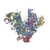
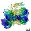
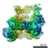
Journal: Nature / Year: 2016 Title: Structure of RNA polymerase I transcribing ribosomal DNA genes. Authors: Simon Neyer / Michael Kunz / Christian Geiss / Merle Hantsche / Victor-Valentin Hodirnau / Anja Seybert / Christoph Engel / Margot P Scheffer / Patrick Cramer / Achilleas S Frangakis / Abstract: RNA polymerase I (Pol I) is a highly processive enzyme that transcribes ribosomal DNA (rDNA) and regulates growth of eukaryotic cells. Crystal structures of free Pol I from the yeast Saccharomyces ...RNA polymerase I (Pol I) is a highly processive enzyme that transcribes ribosomal DNA (rDNA) and regulates growth of eukaryotic cells. Crystal structures of free Pol I from the yeast Saccharomyces cerevisiae have revealed dimers of the enzyme stabilized by a 'connector' element and an expanded cleft containing the active centre in an inactive conformation. The central bridge helix was unfolded and a Pol-I-specific 'expander' element occupied the DNA-template-binding site. The structure of Pol I in its active transcribing conformation has yet to be determined, whereas structures of Pol II and Pol III have been solved with bound DNA template and RNA transcript. Here we report structures of active transcribing Pol I from yeast solved by two different cryo-electron microscopy approaches. A single-particle structure at 3.8 Å resolution reveals a contracted active centre cleft with bound DNA and RNA, and a narrowed pore beneath the active site that no longer holds the RNA-cleavage-stimulating domain of subunit A12.2. A structure at 29 Å resolution that was determined from cryo-electron tomograms of Pol I enzymes transcribing cellular rDNA confirms contraction of the cleft and reveals that incoming and exiting rDNA enclose an angle of around 150°. The structures suggest a model for the regulation of transcription elongation in which contracted and expanded polymerase conformations are associated with active and inactive states, respectively.
History
Deposition
Oct 15, 2016
-
Header (metadata) release
Nov 16, 2016
-
Map release
Nov 23, 2016
-
Update
Nov 23, 2016
-
Current status
Nov 23, 2016
Processing site: PDBe / Status: Released
-
Structure visualization
Movie
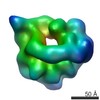
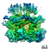
Surface view with section colored by density value
In the structure databanks used in Yorodumi, some data are registered as the other names, "COVID-19 virus" and "2019-nCoV". Here are the details of the virus and the list of structure data.
Jan 31, 2019. EMDB accession codes are about to change! (news from PDBe EMDB page)
EMDB accession codes are about to change! (news from PDBe EMDB page)
The allocation of 4 digits for EMDB accession codes will soon come to an end. Whilst these codes will remain in use, new EMDB accession codes will include an additional digit and will expand incrementally as the available range of codes is exhausted. The current 4-digit format prefixed with “EMD-” (i.e. EMD-XXXX) will advance to a 5-digit format (i.e. EMD-XXXXX), and so on. It is currently estimated that the 4-digit codes will be depleted around Spring 2019, at which point the 5-digit format will come into force.
The EM Navigator/Yorodumi systems omit the EMD- prefix.
Related info.:Q: What is EMD? / ID/Accession-code notation in Yorodumi/EM Navigator
Yorodumi is a browser for structure data from EMDB, PDB, SASBDB, etc.
This page is also the successor to EM Navigator detail page, and also detail information page/front-end page for Omokage search.
The word "yorodu" (or yorozu) is an old Japanese word meaning "ten thousand". "mi" (miru) is to see.
Related info.:EMDB / PDB / SASBDB / Comparison of 3 databanks / Yorodumi Search / Aug 31, 2016. New EM Navigator & Yorodumi / Yorodumi Papers / Jmol/JSmol / Function and homology information / Changes in new EM Navigator and Yorodumi
 Movie
Movie Controller


 Open data
 Basic information
 Map data
 Sample
 Saccharomyces cerevisiae (brewer's yeast)
 Authors
 Citation
 Structure visualization
 Movie viewer
 Downloads & links
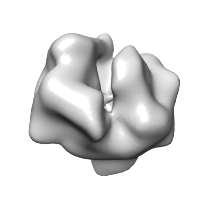 emd_4149.png
 http://ftp.pdbj.org/pub/emdb/structures/EMD-4149


 Sample components
 Processing
 Electron microscopy
