National Institutes of Health/National Institute Of Allergy and Infectious Diseases (NIH/NIAID)
R01 AI127521
United States
Citation
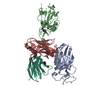
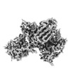
Journal: Commun Biol / Year: 2023 Title: SARS-COV-2 Omicron variants conformationally escape a rare quaternary antibody binding mode. Authors: Jule Goike / Ching-Lin Hsieh / Andrew P Horton / Elizabeth C Gardner / Ling Zhou / Foteini Bartzoka / Nianshuang Wang / Kamyab Javanmardi / Andrew Herbert / Shawn Abbassi / Xuping Xie / ...Authors: Jule Goike / Ching-Lin Hsieh / Andrew P Horton / Elizabeth C Gardner / Ling Zhou / Foteini Bartzoka / Nianshuang Wang / Kamyab Javanmardi / Andrew Herbert / Shawn Abbassi / Xuping Xie / Hongjie Xia / Pei-Yong Shi / Rebecca Renberg / Thomas H Segall-Shapiro / Cynthia I Terrace / Wesley Wu / Raghav Shroff / Michelle Byrom / Andrew D Ellington / Edward M Marcotte / James M Musser / Suresh V Kuchipudi / Vivek Kapur / George Georgiou / Scott C Weaver / John M Dye / Daniel R Boutz / Jason S McLellan / Jimmy D Gollihar / Abstract: The ongoing evolution of SARS-CoV-2 into more easily transmissible and infectious variants has provided unprecedented insight into mutations enabling immune escape. Understanding how these mutations ...The ongoing evolution of SARS-CoV-2 into more easily transmissible and infectious variants has provided unprecedented insight into mutations enabling immune escape. Understanding how these mutations affect the dynamics of antibody-antigen interactions is crucial to the development of broadly protective antibodies and vaccines. Here we report the characterization of a potent neutralizing antibody (N3-1) identified from a COVID-19 patient during the first disease wave. Cryogenic electron microscopy revealed a quaternary binding mode that enables direct interactions with all three receptor-binding domains of the spike protein trimer, resulting in extraordinary avidity and potent neutralization of all major variants of concern until the emergence of Omicron. Structure-based rational design of N3-1 mutants improved binding to all Omicron variants but only partially restored neutralization of the conformationally distinct Omicron BA.1. This study provides new insights into immune evasion through changes in spike protein dynamics and highlights considerations for future conformationally biased multivalent vaccine designs.
In the structure databanks used in Yorodumi, some data are registered as the other names, "COVID-19 virus" and "2019-nCoV". Here are the details of the virus and the list of structure data.
Jan 31, 2019. EMDB accession codes are about to change! (news from PDBe EMDB page)
EMDB accession codes are about to change! (news from PDBe EMDB page)
The allocation of 4 digits for EMDB accession codes will soon come to an end. Whilst these codes will remain in use, new EMDB accession codes will include an additional digit and will expand incrementally as the available range of codes is exhausted. The current 4-digit format prefixed with “EMD-” (i.e. EMD-XXXX) will advance to a 5-digit format (i.e. EMD-XXXXX), and so on. It is currently estimated that the 4-digit codes will be depleted around Spring 2019, at which point the 5-digit format will come into force.
The EM Navigator/Yorodumi systems omit the EMD- prefix.
Related info.:Q: What is EMD? / ID/Accession-code notation in Yorodumi/EM Navigator
Yorodumi is a browser for structure data from EMDB, PDB, SASBDB, etc.
This page is also the successor to EM Navigator detail page, and also detail information page/front-end page for Omokage search.
The word "yorodu" (or yorozu) is an old Japanese word meaning "ten thousand". "mi" (miru) is to see.
Related info.:EMDB / PDB / SASBDB / Comparison of 3 databanks / Yorodumi Search / Aug 31, 2016. New EM Navigator & Yorodumi / Yorodumi Papers / Jmol/JSmol / Function and homology information / Changes in new EM Navigator and Yorodumi
 Movie
Movie Controller


 Open data
 Basic information
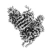
 Map data
 Sample
 Keywords
 neutralizing antibody / RBD-directed antibody / quaternary epitope /
  Function and homology information


 Authors
 United States, 1 items
  Citation
 Structure visualization
 Downloads & links
 emd_41382.png
 http://ftp.pdbj.org/pub/emdb/structures/EMD-41382
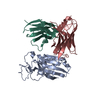


 Z
Z Y
Y X
X


 Sample components
 Processing
 Electron microscopy
