 Movie
Movie Controller


+ Open data

- Basic information
| Entry | Database: EMDB / ID: EMD-4106 | |||||||||
|---|---|---|---|---|---|---|---|---|---|---|
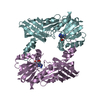
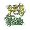
| Title | TRF1-DNA | |||||||||
 Map data Map data | TRF1 bound to seven repeats of telomeric dsDNA | |||||||||
 Sample Sample |
| |||||||||
| Biological species |  | |||||||||
| Method | single particle reconstruction / negative staining / Resolution: 25.0 Å | |||||||||
 Authors Authors | Boskovic J / Martinez-Gago J / Mendez-Pertuz M / Buscato A / Martinez-Torrecuadrada JL / Blasco MA | |||||||||
 Citation Citation | Journal: J Biol Chem / Year: 2016 Title: Molecular Architecture of Full-length TRF1 Favors Its Interaction with DNA. Authors: Jasminka Boskovic / Jaime Martinez-Gago / Marinela Mendez-Pertuz / Alberto Buscato / Jorge Luis Martinez-Torrecuadrada / Maria A Blasco /  Abstract: Telomeres are specific DNA-protein structures found at both ends of eukaryotic chromosomes that protect the genome from degradation and from being recognized as double-stranded breaks. In ...Telomeres are specific DNA-protein structures found at both ends of eukaryotic chromosomes that protect the genome from degradation and from being recognized as double-stranded breaks. In vertebrates, telomeres are composed of tandem repeats of the TTAGGG sequence that are bound by a six-subunit complex called shelterin. Molecular mechanisms of telomere functions remain unknown in large part due to lack of structural data on shelterins, shelterin complex, and its interaction with the telomeric DNA repeats. TRF1 is one of the best studied shelterin components; however, the molecular architecture of the full-length protein remains unknown. We have used single-particle electron microscopy to elucidate the structure of TRF1 and its interaction with telomeric DNA sequence. Our results demonstrate that full-length TRF1 presents a molecular architecture that assists its interaction with telometic DNA and at the same time makes TRFH domains accessible to other TRF1 binding partners. Furthermore, our studies suggest hypothetical models on how other proteins as TIN2 and tankyrase contribute to regulate TRF1 function. | |||||||||
| History |
|
- Structure visualization
| Movie |
 Movie viewer Movie viewer |
|---|---|
| Structure viewer | EM map: SurfViewMolmilJmol/JSmol |
| Supplemental images |
 UCSF Chimera
- Downloads & links
-EMDB archive
| Map data | emd_4106.map.gz | 1.7 MB | EMDB map data format | |
|---|---|---|---|---|
| Header (meta data) | emd-4106-v30.xmlemd-4106.xml | 12.1 KB 12.1 KB | Display Display | EMDB header |
| Images | 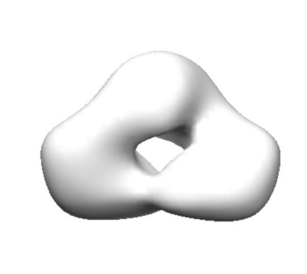 emd_4106.png emd_4106.png | 30 KB | ||
| Archive directory |  http://ftp.pdbj.org/pub/emdb/structures/EMD-4106ftp://ftp.pdbj.org/pub/emdb/structures/EMD-4106 http://ftp.pdbj.org/pub/emdb/structures/EMD-4106ftp://ftp.pdbj.org/pub/emdb/structures/EMD-4106 | HTTPS FTP |
-Validation report
| Summary document | emd_4106_validation.pdf.gz | 189.1 KB | Display | EMDB validaton report |
|---|---|---|---|---|
| Full document | emd_4106_full_validation.pdf.gz | 188.2 KB | Display | |
| Data in XML | emd_4106_validation.xml.gz | 4.8 KB | Display | |
| Arichive directory | https://ftp.pdbj.org/pub/emdb/validation_reports/EMD-4106ftp://ftp.pdbj.org/pub/emdb/validation_reports/EMD-4106 | HTTPS FTP |
-Related structure data


-Links
| EMDB pages | EMDB (EBI/PDBe) / EMDataResource |
|---|
-Map
| File | Download / File: emd_4106.map.gz / Format: CCP4 / Size: 2 MB / Type: IMAGE STORED AS FLOATING POINT NUMBER (4 BYTES) | ||||||||||||||||||||||||||||||||||||||||||||||||||||||||||||
|---|---|---|---|---|---|---|---|---|---|---|---|---|---|---|---|---|---|---|---|---|---|---|---|---|---|---|---|---|---|---|---|---|---|---|---|---|---|---|---|---|---|---|---|---|---|---|---|---|---|---|---|---|---|---|---|---|---|---|---|---|---|
| Annotation | TRF1 bound to seven repeats of telomeric dsDNA | ||||||||||||||||||||||||||||||||||||||||||||||||||||||||||||
| Voxel size | X=Y=Z: 2.5 Å | ||||||||||||||||||||||||||||||||||||||||||||||||||||||||||||
| Density |
| ||||||||||||||||||||||||||||||||||||||||||||||||||||||||||||
| Symmetry | Space group: 1 | ||||||||||||||||||||||||||||||||||||||||||||||||||||||||||||
| Details | EMDB XML:
CCP4 map header:
| ||||||||||||||||||||||||||||||||||||||||||||||||||||||||||||
-Supplemental data
- Sample components
-Entire : TRF1-DNA complex
| Entire | Name: TRF1-DNA complex |
|---|---|
| Components |
|
-Supramolecule #1: TRF1-DNA complex
| Supramolecule | Name: TRF1-DNA complex / type: complex / ID: 1 / Parent: 0 Details: TRF1 protein bound to seven repeats (TTAGGG) of telomeric dsDNA |
|---|---|
| Source (natural) | Organism: |
| Recombinant expression | Organism:   Spodoptera frugiperda (fall armyworm) / Recombinant strain: Sf9 / Recombinant plasmid: pBacPak8 Spodoptera frugiperda (fall armyworm) / Recombinant strain: Sf9 / Recombinant plasmid: pBacPak8 |
| Molecular weight | Experimental: 100 KDa |
-Experimental details
-Structure determination
| Method | negative staining |
|---|---|
 Processing Processing | single particle reconstruction |
| Aggregation state | particle |
-Sample preparation
| Concentration | 0.08 mg/mL | ||||||||||||
|---|---|---|---|---|---|---|---|---|---|---|---|---|---|
| Buffer | pH: 7.5 Component:
| ||||||||||||
| Staining | Type: NEGATIVE / Material: Uranyl Acetate Details: Negatively stained EM specimens were prepared using standar negative staining technique with 1% UrAc | ||||||||||||
| Grid | Model: Electron Microscopy Science / Material: COPPER / Mesh: 400 / Support film - Material: CARBON / Support film - topology: CONTINUOUS / Pretreatment - Type: GLOW DISCHARGE / Pretreatment - Atmosphere: AIR |
- Electron microscopy
| Microscope | FEI TECNAI 12 |
|---|---|
| Image recording | Film or detector model: TVIPS TEMCAM-F416 (4k x 4k) / Digitization - Sampling interval: 15.6 µm / Number grids imaged: 1 / Number real images: 397 / Average exposure time: 1.0 sec. / Average electron dose: 15.0 e/Å2 |
| Electron beam | Acceleration voltage: 120 kV / Electron source: LAB6 |
| Electron optics | Illumination mode: FLOOD BEAM / Imaging mode: BRIGHT FIELD / Cs: 2.2 mm / Nominal defocus max: 3.5 µm / Nominal magnification: 61350 |
| Sample stage | Specimen holder model: SIDE ENTRY, EUCENTRIC |
+Image processing
-Atomic model buiding 1
| Refinement | Space: REAL / Protocol: RIGID BODY FIT / Target criteria: visual inspection |
|---|
