 Movie
Movie Controller


+ Open data

- Basic information
| Entry |  | |||||||||
|---|---|---|---|---|---|---|---|---|---|---|
| Title | Cryo-EM structure of a full-length, native Drp1 dimer | |||||||||
 Map data Map data | Drp1 cryoEM native dimer map | |||||||||
 Sample Sample |
| |||||||||
 Keywords Keywords | Dynamin-related protein 1 /  Drp1 / mitochondrial fission / GTPase / Cytosolic Protein Drp1 / mitochondrial fission / GTPase / Cytosolic Protein | |||||||||
| Function / homology |  Function and homology information Function and homology informationmitochondrial membrane fission / regulation of ATP metabolic process / regulation of peroxisome organization / mitocytosis / Apoptotic execution phase / dynamin GTPase / peroxisome fission / regulation of mitophagy / mitochondrial fragmentation involved in apoptotic process / GTP-dependent protein binding ...mitochondrial membrane fission / regulation of ATP metabolic process / regulation of peroxisome organization / mitocytosis / Apoptotic execution phase / dynamin GTPase / peroxisome fission / regulation of mitophagy / mitochondrial fragmentation involved in apoptotic process / GTP-dependent protein binding / protein localization to mitochondrion / mitochondrial fission / positive regulation of neutrophil chemotaxis / regulation of mitochondrion organization / positive regulation of mitochondrial fission / intracellular distribution of mitochondria / heart contraction / necroptotic process / positive regulation of release of cytochrome c from mitochondria / brush border / localization / positive regulation of intrinsic apoptotic signaling pathway / clathrin-coated pit / GTPase activator activity / mitochondrion organization / release of cytochrome c from mitochondria / positive regulation of protein secretion / synaptic vesicle membrane / small GTPase binding / peroxisome / endocytosis / calcium ion transport / rhythmic process / protein complex oligomerization / microtubule binding / regulation of gene expression / protein-containing complex assembly / microtubule / mitochondrial outer membrane / membrane fusion / molecular adaptor activity / positive regulation of apoptotic process / intracellular membrane-bounded organelle / GTPase activity / lipid binding / ubiquitin protein ligase binding / endoplasmic reticulum membrane / GTP binding / perinuclear region of cytoplasm / Golgi apparatus / endoplasmic reticulum / protein homodimerization activity / protein-containing complex / mitochondrion / membrane / identical protein binding / cytosol / cytoplasmSimilarity search - Function | |||||||||
| Biological species |  Homo sapiens (human) Homo sapiens (human) | |||||||||
| Method | single particle reconstruction / cryo EM / Resolution: 5.97 Å | |||||||||
 Authors Authors | Rochon K / Mears JA | |||||||||
| Funding support |  United States, 2 items United States, 2 items
| |||||||||
 Citation Citation | Journal: Nat Commun / Year: 2024 Title: Structural basis for regulated assembly of the mitochondrial fission GTPase Drp1. Authors: Kristy Rochon / Brianna L Bauer / Nathaniel A Roethler / Yuli Buckley / Chih-Chia Su / Wei Huang / Rajesh Ramachandran / Maria S K Stoll / Edward W Yu / Derek J Taylor / Jason A Mears / Abstract: Mitochondrial fission is a critical cellular event to maintain organelle function. This multistep process is initiated by the enhanced recruitment and oligomerization of dynamin-related protein 1 ...Mitochondrial fission is a critical cellular event to maintain organelle function. This multistep process is initiated by the enhanced recruitment and oligomerization of dynamin-related protein 1 (Drp1) at the surface of mitochondria. As such, Drp1 is essential for inducing mitochondrial division in mammalian cells, and homologous proteins are found in all eukaryotes. As a member of the dynamin superfamily of proteins (DSPs), controlled Drp1 self-assembly into large helical polymers stimulates its GTPase activity to promote membrane constriction. Still, little is known about the mechanisms that regulate correct spatial and temporal assembly of the fission machinery. Here we present a cryo-EM structure of a full-length Drp1 dimer in an auto-inhibited state. This dimer reveals two key conformational rearrangements that must be unlocked through intramolecular rearrangements to achieve the assembly-competent state observed in previous structures. This structural insight provides understanding into the mechanism for regulated self-assembly of the mitochondrial fission machinery. | |||||||||
| History |
|
- Structure visualization
| Supplemental images |
|---|

- Downloads & links
-EMDB archive
| Map data | emd_40967.map.gz | 156.7 MB | EMDB map data format | |
|---|---|---|---|---|
| Header (meta data) | emd-40967-v30.xmlemd-40967.xml | 18.1 KB 18.1 KB | Display Display | EMDB header |
| FSC (resolution estimation) | emd_40967_fsc.xml | 11.7 KB | Display | FSC data file |
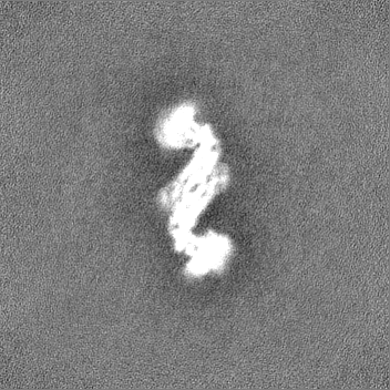
| Images |  emd_40967.png emd_40967.png | 47.9 KB | ||
| Filedesc metadata | emd-40967.cif.gz | 6.5 KB | ||
| Others | emd_40967_half_map_1.map.gzemd_40967_half_map_2.map.gz | 154.2 MB 154.2 MB | ||
| Archive directory |  http://ftp.pdbj.org/pub/emdb/structures/EMD-40967ftp://ftp.pdbj.org/pub/emdb/structures/EMD-40967 http://ftp.pdbj.org/pub/emdb/structures/EMD-40967ftp://ftp.pdbj.org/pub/emdb/structures/EMD-40967 | HTTPS FTP |
-Related structure data
| Related structure data |  8t1hMC M: atomic model generated by this map C: citing same article ( |
|---|---|
| Similar structure data |
-Links
| EMDB pages | EMDB (EBI/PDBe) / EMDataResource |
|---|
-Map
| File | Download / File: emd_40967.map.gz / Format: CCP4 / Size: 166.4 MB / Type: IMAGE STORED AS FLOATING POINT NUMBER (4 BYTES) | ||||||||||||||||||||||||||||||||||||
|---|---|---|---|---|---|---|---|---|---|---|---|---|---|---|---|---|---|---|---|---|---|---|---|---|---|---|---|---|---|---|---|---|---|---|---|---|---|
| Annotation | Drp1 cryoEM native dimer map | ||||||||||||||||||||||||||||||||||||
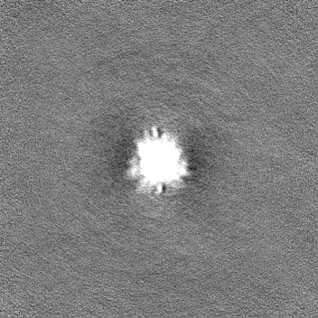
| Projections & slices | Image control
Images are generated by Spider. | ||||||||||||||||||||||||||||||||||||
| Voxel size | X=Y=Z: 1.07 Å | ||||||||||||||||||||||||||||||||||||
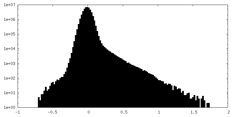
| Density |
| ||||||||||||||||||||||||||||||||||||
| Symmetry | Space group: 1 | ||||||||||||||||||||||||||||||||||||
| Details | EMDB XML:
|
 Z (Sec.)
 Y (Row.)
 X (Col.)


-Supplemental data
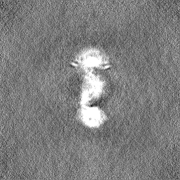
-Half map: Drp1 cryoEM native dimer half map B
| File | emd_40967_half_map_1.map | ||||||||||||
|---|---|---|---|---|---|---|---|---|---|---|---|---|---|
| Annotation | Drp1 cryoEM native dimer half map B | ||||||||||||
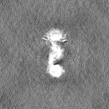
| Projections & Slices |
| ||||||||||||
| Density Histograms |


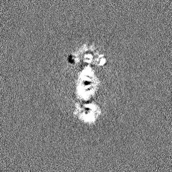

-Half map: Drp1 cryoEM native dimer half map A
| File | emd_40967_half_map_2.map | ||||||||||||
|---|---|---|---|---|---|---|---|---|---|---|---|---|---|
| Annotation | Drp1 cryoEM native dimer half map A | ||||||||||||
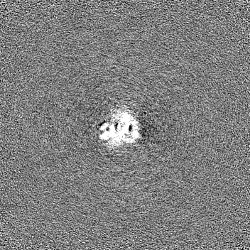
| Projections & Slices |
| ||||||||||||
| Density Histograms |


- Sample components
-Entire : Dimer complex of native, full length Drp1
| Entire | Name: Dimer complex of native, full length Drp1 |
|---|---|
| Components |
|
-Supramolecule #1: Dimer complex of native, full length Drp1
| Supramolecule | Name: Dimer complex of native, full length Drp1 / type: complex / ID: 1 / Parent: 0 / Macromolecule list: all |
|---|---|
| Source (natural) | Organism: Homo sapiens (human) |
| Molecular weight | Theoretical: 164 KDa |
-Macromolecule #1: Dynamin-1-like protein
| Macromolecule | Name: Dynamin-1-like protein / type: protein_or_peptide / ID: 1 / Number of copies: 2 / Enantiomer: LEVO / EC number: dynamin GTPase |
|---|---|
| Source (natural) | Organism: Homo sapiens (human) |
| Molecular weight | Theoretical: 81.984094 KDa |
| Recombinant expression | Organism:  Escherichia coli (E. coli) Escherichia coli (E. coli) |
| Sequence | String: MEALIPVINK LQDVFNTVGA DIIQLPQIVV VGTQSSGKSS VLESLVGRDL LPRGTGIVTR RPLILQLVHV SQEDKRKTTG EENGVEAEE WGKFLHTKNK LYTDFDEIRQ EIENETERIS GNNKGVSPEP IHLKIFSPNV VNLTLVDLPG MTKVPVGDQP K DIELQIRE ...String: MEALIPVINK LQDVFNTVGA DIIQLPQIVV VGTQSSGKSS VLESLVGRDL LPRGTGIVTR RPLILQLVHV SQEDKRKTTG EENGVEAEE WGKFLHTKNK LYTDFDEIRQ EIENETERIS GNNKGVSPEP IHLKIFSPNV VNLTLVDLPG MTKVPVGDQP K DIELQIRE LILRFISNPN SIILAVTAAN TDMATSEALK ISREVDPDGR RTLAVITKLD LMDAGTDAMD VLMGRVIPVK LG IIGVVNR SQLDINNKKS VTDSIRDEYA FLQKKYPSLA NRNGTKYLAR TLNRLLMHHI RDCLPELKTR INVLAAQYQS LLN SYGEPV DDKSATLLQL ITKFATEYCN TIEGTAKYIE TSELCGGARI CYIFHETFGR TLESVDPLGG LNTIDILTAI RNAT GPRPA LFVPEVSFEL LVKRQIKRLE EPSLRCVELV HEEMQRIIQH CSNYSTQELL RFPKLHDAIV EVVTCLLRKR LPVTN EMVH NLVAIELAYI NTKHPDFADA CGLMNNNIEE QRRNRLAREL PSAVSRDKSS KVPSALAPAS QEPSPAASAE ADGKLI QDS RRETKNVASG GGGVGDGVQE PTTGNWRGML KTSKAEELLA EEKSKPIPIM PASPQKGHAV NLLDVPVPVA RKLSARE QR DCEVIERLIK SYFLIVRKNI QDSVPKAVMH FLVNHVKDTL QSELVGQLYK SSLLDDLLTE SEDMAQRRKE AADMLKAL Q GASQIIAEIR ETHLW UniProtKB: Dynamin-1-like protein |
-Experimental details
-Structure determination
| Method | cryo EM |
|---|---|
 Processing Processing | single particle reconstruction |
| Aggregation state | particle |
-Sample preparation
| Concentration | .05 mg/mL | |||||||||||||||
|---|---|---|---|---|---|---|---|---|---|---|---|---|---|---|---|---|
| Buffer | pH: 7.5 Component:
Details: 25 mM HEPES (KOH) pH 7.5, 0.15 M KCl, 5 mM MgCl2, 10 mM Beta-mercaptoethanol | |||||||||||||||
| Grid | Model: Quantifoil R1.2/1.3 / Material: COPPER / Mesh: 300 / Support film - Material: GRAPHENE OXIDE / Support film - topology: CONTINUOUS | |||||||||||||||
| Vitrification | Cryogen name: ETHANE / Chamber humidity: 100 % / Chamber temperature: 277.15 K / Instrument: FEI VITROBOT MARK III |
- Electron microscopy
| Microscope | FEI TITAN KRIOS |
|---|---|
| Electron beam | Acceleration voltage: 300 kV / Electron source: FIELD EMISSION GUN |
| Electron optics | Illumination mode: SPOT SCAN / Imaging mode: BRIGHT FIELDBright-field microscopy / Nominal defocus max: 2.0 µm / Nominal defocus min: 0.8 µm |
| Sample stage | Cooling holder cryogen: NITROGEN |
| Image recording | Film or detector model: GATAN K2 SUMMIT (4k x 4k) / Detector mode: SUPER-RESOLUTION / Number real images: 4560 / Average electron dose: 47.76 e/Å2 |
| Experimental equipment |  Model: Titan Krios / Image courtesy: FEI Company |
-Image processing
| Particle selection | Number selected: 883690 |
|---|---|
| Startup model | Type of model: NONE |
| Initial angle assignment | Type: MAXIMUM LIKELIHOOD / Software - Name: cryoSPARC (ver. 3) |
| Final 3D classification | Number classes: 4 / Software - Name: cryoSPARC (ver. 3) |
| Final angle assignment | Type: MAXIMUM LIKELIHOOD / Software - Name: cryoSPARC (ver. 3) |
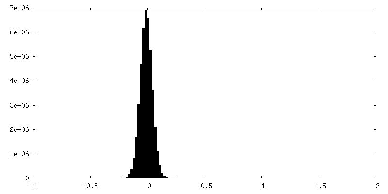
| Final reconstruction | Applied symmetry - Point group: C1 (asymmetric) / Resolution.type: BY AUTHOR / Resolution: 5.97 Å / Resolution method: FSC 0.143 CUT-OFF / Software - Name: cryoSPARC (ver. 3) / Number images used: 71611 |
| FSC plot (resolution estimation) |  |

