 Movie
Movie Controller

[English] 日本語
 Yorodumi
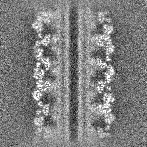
- EMDB-40903: Cryo-EM of the GDP-bound human dynamin polymer assembled on the m... -
+ Open data

- Basic information
| Entry |  | ||||||||||||
|---|---|---|---|---|---|---|---|---|---|---|---|---|---|
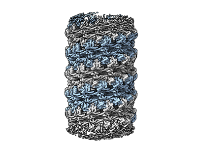
| Title | Cryo-EM of the GDP-bound human dynamin polymer assembled on the membrane in the super constricted state (full helix) | ||||||||||||
 Map data Map data | |||||||||||||
 Sample Sample |
| ||||||||||||
 Keywords Keywords |  dynamin / membrane / fission / lipid / tubule / scission / endocytosis / HYDROLASE dynamin / membrane / fission / lipid / tubule / scission / endocytosis / HYDROLASE | ||||||||||||
| Function / homology |  Function and homology information Function and homology informationclathrin coat assembly involved in endocytosis / vesicle scission / synaptic vesicle budding from presynaptic endocytic zone membrane / presynaptic endocytic zone membrane / dynamin GTPase / chromaffin granule / regulation of vesicle size / photoreceptor ribbon synapse / Toll Like Receptor 4 (TLR4) Cascade / Retrograde neurotrophin signalling ...clathrin coat assembly involved in endocytosis / vesicle scission / synaptic vesicle budding from presynaptic endocytic zone membrane / presynaptic endocytic zone membrane / dynamin GTPase / chromaffin granule / regulation of vesicle size / photoreceptor ribbon synapse / Toll Like Receptor 4 (TLR4) Cascade / Retrograde neurotrophin signalling / Formation of annular gap junctions / endosome organization / Gap junction degradation / membrane coat / Recycling pathway of L1 / phosphatidylinositol-3,4,5-trisphosphate binding / synaptic vesicle endocytosis / endocytic vesicle / EPH-ephrin mediated repulsion of cells / clathrin-coated pit / phosphatidylinositol-4,5-bisphosphate binding / MHC class II antigen presentation / photoreceptor inner segment / receptor-mediated endocytosis / cell projection / modulation of chemical synaptic transmission / protein homooligomerization / receptor internalization / endocytosis / GDP binding / : / presynapse / Clathrin-mediated endocytosis / microtubule binding / protein homotetramerization / microtubule / GTPase activity / glutamatergic synapse / GTP binding / protein kinase binding / protein homodimerization activity / RNA binding / extracellular exosome / membrane / identical protein binding / plasma membrane / cytoplasmSimilarity search - Function | ||||||||||||
| Biological species |  Homo sapiens (human) Homo sapiens (human) | ||||||||||||
| Method | helical reconstruction / cryo EM / Resolution: 3.55 Å | ||||||||||||
 Authors Authors | Jimah JR / Canagarajah BJ / Hinshaw JE | ||||||||||||
| Funding support |  United States, 3 items United States, 3 items
| ||||||||||||
 Citation Citation | Journal: Dev Cell / Year: 2024 Title: Cryo-EM structures of membrane-bound dynamin in a post-hydrolysis state primed for membrane fission. Authors: John R Jimah / Nidhi Kundu / Abigail E Stanton / Kem A Sochacki / Bertram Canagarajah / Lieza Chan / Marie-Paule Strub / Huaibin Wang / Justin W Taraska / Jenny E Hinshaw / Abstract: Dynamin assembles as a helical polymer at the neck of budding endocytic vesicles, constricting the underlying membrane as it progresses through the GTPase cycle to sever vesicles from the ...Dynamin assembles as a helical polymer at the neck of budding endocytic vesicles, constricting the underlying membrane as it progresses through the GTPase cycle to sever vesicles from the plasma membrane. Although atomic models of the dynamin helical polymer bound to guanosine triphosphate (GTP) analogs define earlier stages of membrane constriction, there are no atomic models of the assembled state post-GTP hydrolysis. Here, we used cryo-EM methods to determine atomic structures of the dynamin helical polymer assembled on lipid tubules, akin to necks of budding endocytic vesicles, in a guanosine diphosphate (GDP)-bound, super-constricted state. In this state, dynamin is assembled as a 2-start helix with an inner lumen of 3.4 nm, primed for spontaneous fission. Additionally, by cryo-electron tomography, we trapped dynamin helical assemblies within HeLa cells using the GTPase-defective dynamin K44A mutant and observed diverse dynamin helices, demonstrating that dynamin can accommodate a range of assembled complexes in cells that likely precede membrane fission. | ||||||||||||
| History |
|
- Structure visualization
| Supplemental images |
|---|
- Downloads & links
-EMDB archive
| Map data | emd_40903.map.gz | 57.7 MB | EMDB map data format | |
|---|---|---|---|---|
| Header (meta data) | emd-40903-v30.xmlemd-40903.xml | 15.8 KB 15.8 KB | Display Display | EMDB header |
| FSC (resolution estimation) | emd_40903_fsc.xml | 18.6 KB | Display | FSC data file |
| Images |  emd_40903.png emd_40903.png | 99.9 KB | ||
| Masks | emd_40903_msk_1.map | 476.8 MB | Mask map | |
| Filedesc metadata | emd-40903.cif.gz | 6 KB | ||
| Others | emd_40903_half_map_1.map.gzemd_40903_half_map_2.map.gz | 442 MB 442 MB | ||
| Archive directory |  http://ftp.pdbj.org/pub/emdb/structures/EMD-40903ftp://ftp.pdbj.org/pub/emdb/structures/EMD-40903 http://ftp.pdbj.org/pub/emdb/structures/EMD-40903ftp://ftp.pdbj.org/pub/emdb/structures/EMD-40903 | HTTPS FTP |
-Related structure data
| Related structure data |  8sz8MC  8sxzC  8sz4C 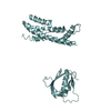 8sz7C  8t0kC  8t0rC C: citing same article ( M: atomic model generated by this map |
|---|---|
| Similar structure data |


-Links
| EMDB pages | EMDB (EBI/PDBe) / EMDataResource |
|---|---|
| Related items in Molecule of the Month |


-Map
| File | Download / File: emd_40903.map.gz / Format: CCP4 / Size: 476.8 MB / Type: IMAGE STORED AS FLOATING POINT NUMBER (4 BYTES) | ||||||||||||||||||||
|---|---|---|---|---|---|---|---|---|---|---|---|---|---|---|---|---|---|---|---|---|---|
| Voxel size | X=Y=Z: 1.289 Å | ||||||||||||||||||||
| Density |
| ||||||||||||||||||||
| Symmetry | Space group: 1 | ||||||||||||||||||||
| Details | EMDB XML:
|
-Supplemental data
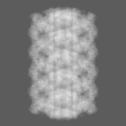
-Mask #1
| File | emd_40903_msk_1.map | ||||||||||||
|---|---|---|---|---|---|---|---|---|---|---|---|---|---|
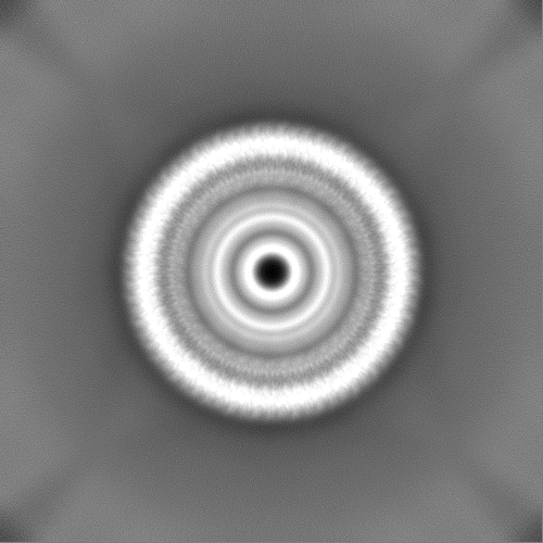
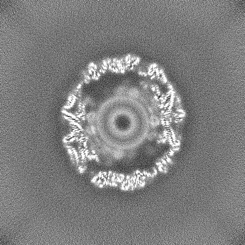
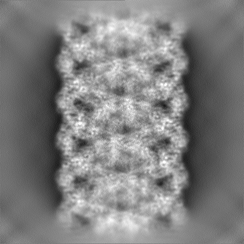
| Projections & Slices |
| ||||||||||||
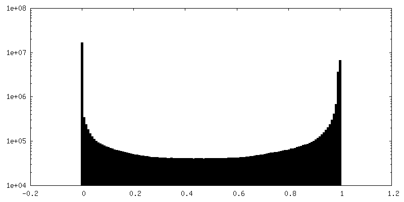
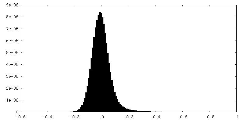
| Density Histograms |
 Z
Z Y
Y X
X


-Half map: #1
| File | emd_40903_half_map_1.map | ||||||||||||
|---|---|---|---|---|---|---|---|---|---|---|---|---|---|
| Projections & Slices |
| ||||||||||||
| Density Histograms |


-Half map: #2
| File | emd_40903_half_map_2.map | ||||||||||||
|---|---|---|---|---|---|---|---|---|---|---|---|---|---|
| Projections & Slices |
| ||||||||||||
| Density Histograms |


- Sample components
-Entire : Cryo-EM of the GDP-bound human dynamin polymer assembled on the m...
| Entire | Name: Cryo-EM of the GDP-bound human dynamin polymer assembled on the membrane in the super constricted state (full helix) |
|---|---|
| Components |
|
-Supramolecule #1: Cryo-EM of the GDP-bound human dynamin polymer assembled on the m...
| Supramolecule | Name: Cryo-EM of the GDP-bound human dynamin polymer assembled on the membrane in the super constricted state (full helix) type: complex / ID: 1 / Parent: 0 / Macromolecule list: #1 |
|---|---|
| Source (natural) | Organism: Homo sapiens (human) |
| Molecular weight | Theoretical: 85.7 kDa/nm |
-Macromolecule #1: Dynamin-1
| Macromolecule | Name: Dynamin-1 / type: protein_or_peptide / ID: 1 / Number of copies: 4 / Enantiomer: LEVO / EC number: dynamin GTPase |
|---|---|
| Source (natural) | Organism: Homo sapiens (human) |
| Molecular weight | Theoretical: 86.312625 KDa |
| Recombinant expression | Organism:  Escherichia coli 'BL21-Gold(DE3)pLysS AG' (bacteria) Escherichia coli 'BL21-Gold(DE3)pLysS AG' (bacteria) |
| Sequence | String: GPTGMGNRGM EDLIPLVNRL QDAFSAIGQN ADLDLPQIAV VGGQSAGASS VLENFVGRDF LPRGSGIVTR RPLVLQLVNA TTEYAEFLH CKGKKFTDFE EVRLEIEAET DRVTGTNKGI SPVPINLRVY SPHVLNLTLV DLPGMTKVPV GDQPPDIEFQ I RDMLMQFV ...String: GPTGMGNRGM EDLIPLVNRL QDAFSAIGQN ADLDLPQIAV VGGQSAGASS VLENFVGRDF LPRGSGIVTR RPLVLQLVNA TTEYAEFLH CKGKKFTDFE EVRLEIEAET DRVTGTNKGI SPVPINLRVY SPHVLNLTLV DLPGMTKVPV GDQPPDIEFQ I RDMLMQFV TKENCLILAV SPANSDLANS DALKVAKEVD PQGQRTIGVI TKLDLMDEGT DARDVLENKL LPLRRGYIGV VN RSQKDID GKKDITAALA AERKFFLSHP SYRHLADRMG TPYLQKVLNQ QLTNHIRDTL PGLRNKLQSQ LLSIEKEVEE YKN FRPDDP ARKTKALLQM VQQFAVDFEK RIEGSGDQID TYELSGGARI NRIFHERFPF ELVKMEFDEK ELRREISYAI KNIH GIRTG LFTPDMAFET IVKKQVKKIR EPCLKCVDMV ISELISTVRQ CTKKLQQYPR LREEMERIVT THIREREGRT KEQVM LLID IELAYMNTNH EDFIGFANAQ QRSNQMNKKK TSGNQDEILV IRKGWLTINN IGIMKGGSKE YWFVLTAENL SWYKDD EEK EKKYMLSVDN LKLRDVEKGF MSSKHIFALF NTEQRNVYKD YRQLELACET QEEVDSWKAS FLRAGVYPER VGDKEKA SE TEENGSDSFM HSMDPQLERQ VETIRNLVDS YMAIVNKTVR DLMPKTIMHL MINNTKEFIF SELLANLYSC GDQNTLME E SAEQAQRRDE MLRMYHALKE ALSIIGNINT TTV UniProtKB: Dynamin-1 |
-Macromolecule #2: GUANOSINE-5'-DIPHOSPHATE
| Macromolecule | Name: GUANOSINE-5'-DIPHOSPHATE / type: ligand / ID: 2 / Number of copies: 4 / Formula: GDP |
|---|---|
| Molecular weight | Theoretical: 443.201 Da |
| Chemical component information |  ChemComp-GDP: |
-Macromolecule #3: MAGNESIUM ION
| Macromolecule | Name: MAGNESIUM ION / type: ligand / ID: 3 / Number of copies: 4 / Formula: MG |
|---|---|
| Molecular weight | Theoretical: 24.305 Da |
-Experimental details
-Structure determination
| Method | cryo EM |
|---|---|
 Processing Processing | helical reconstruction |
| Aggregation state | filament |
-Sample preparation
| Buffer | pH: 7.4 |
|---|---|
| Vitrification | Cryogen name: ETHANE |
- Electron microscopy
| Microscope | TFS KRIOS |
|---|---|
| Electron beam | Acceleration voltage: 300 kV / Electron source: FIELD EMISSION GUN |
| Electron optics | Illumination mode: FLOOD BEAM / Imaging mode: BRIGHT FIELDBright-field microscopy / Nominal defocus max: 1.8 µm / Nominal defocus min: 0.6 µm |
| Image recording | Film or detector model: GATAN K2 SUMMIT (4k x 4k) / Average electron dose: 60.0 e/Å2 |
| Experimental equipment |  Model: Titan Krios / Image courtesy: FEI Company |
-Image processing
| Startup model | Type of model: OTHER |
|---|---|
| Final angle assignment | Type: NOT APPLICABLE |
| Final reconstruction | Applied symmetry - Helical parameters - Δz: 14.675 Å Applied symmetry - Helical parameters - Δ&Phi: 26.146 ° Applied symmetry - Helical parameters - Axial symmetry: C1 (asymmetric) Resolution.type: BY AUTHOR / Resolution: 3.55 Å / Resolution method: FSC 0.143 CUT-OFF / Number images used: 609714 |
| FSC plot (resolution estimation) |  |
