Movie
Movie Controller

[English] 日本語
 Yorodumi
- EMDB-40557: Cryo-EM structure of designed Influenza HA binder, HA_20, bound t... -
+ Open data

- Basic information
| Entry |  | |||||||||
|---|---|---|---|---|---|---|---|---|---|---|
| Title | Cryo-EM structure of designed Influenza HA binder, HA_20, bound to Influenza HA (Strain: Iowa43) | |||||||||
 Map data Map data | unsharpened | |||||||||
 Sample Sample |
| |||||||||
 Keywords Keywords | flu /  influenza / hemagglutinin / HA / Iowa43 / HA_20 / DE NOVO PROTEIN / minibinder / binder / designed protein / fusion protein / glycoprotein / DE NOVO PROTEIN-Viral Protein complex influenza / hemagglutinin / HA / Iowa43 / HA_20 / DE NOVO PROTEIN / minibinder / binder / designed protein / fusion protein / glycoprotein / DE NOVO PROTEIN-Viral Protein complex | |||||||||
| Function / homology |  Function and homology informationviral budding from plasma membrane / clathrin-dependent endocytosis of virus by host cell / host cell surface receptor binding / fusion of virus membrane with host plasma membrane / fusion of virus membrane with host endosome membrane / viral envelope / virion attachment to host cell / host cell plasma membrane / virion membrane / membrane Function and homology informationviral budding from plasma membrane / clathrin-dependent endocytosis of virus by host cell / host cell surface receptor binding / fusion of virus membrane with host plasma membrane / fusion of virus membrane with host endosome membrane / viral envelope / virion attachment to host cell / host cell plasma membrane / virion membrane / membraneSimilarity search - Function | |||||||||
| Biological species |   Influenza A virus / unidentified (others) Influenza A virus / unidentified (others) | |||||||||
| Method | single particle reconstruction / cryo EM / Resolution: 2.93 Å | |||||||||
 Authors Authors | Borst AJ / Bennett NR | |||||||||
| Funding support |  United States, 1 items United States, 1 items
| |||||||||
 Citation Citation | Journal: Nature / Year: 2023 Title: De novo design of protein structure and function with RFdiffusion. Authors: Joseph L Watson / David Juergens / Nathaniel R Bennett / Brian L Trippe / Jason Yim / Helen E Eisenach / Woody Ahern / Andrew J Borst / Robert J Ragotte / Lukas F Milles / Basile I M Wicky / ...Authors: Joseph L Watson / David Juergens / Nathaniel R Bennett / Brian L Trippe / Jason Yim / Helen E Eisenach / Woody Ahern / Andrew J Borst / Robert J Ragotte / Lukas F Milles / Basile I M Wicky / Nikita Hanikel / Samuel J Pellock / Alexis Courbet / William Sheffler / Jue Wang / Preetham Venkatesh / Isaac Sappington / Susana Vázquez Torres / Anna Lauko / Valentin De Bortoli / Emile Mathieu / Sergey Ovchinnikov / Regina Barzilay / Tommi S Jaakkola / Frank DiMaio / Minkyung Baek / David Baker /    Abstract: There has been considerable recent progress in designing new proteins using deep-learning methods. Despite this progress, a general deep-learning framework for protein design that enables solution of ...There has been considerable recent progress in designing new proteins using deep-learning methods. Despite this progress, a general deep-learning framework for protein design that enables solution of a wide range of design challenges, including de novo binder design and design of higher-order symmetric architectures, has yet to be described. Diffusion models have had considerable success in image and language generative modelling but limited success when applied to protein modelling, probably due to the complexity of protein backbone geometry and sequence-structure relationships. Here we show that by fine-tuning the RoseTTAFold structure prediction network on protein structure denoising tasks, we obtain a generative model of protein backbones that achieves outstanding performance on unconditional and topology-constrained protein monomer design, protein binder design, symmetric oligomer design, enzyme active site scaffolding and symmetric motif scaffolding for therapeutic and metal-binding protein design. We demonstrate the power and generality of the method, called RoseTTAFold diffusion (RFdiffusion), by experimentally characterizing the structures and functions of hundreds of designed symmetric assemblies, metal-binding proteins and protein binders. The accuracy of RFdiffusion is confirmed by the cryogenic electron microscopy structure of a designed binder in complex with influenza haemagglutinin that is nearly identical to the design model. In a manner analogous to networks that produce images from user-specified inputs, RFdiffusion enables the design of diverse functional proteins from simple molecular specifications. | |||||||||
| History |
|
- Structure visualization
| Supplemental images |
|---|

- Downloads & links
-EMDB archive
| Map data | emd_40557.map.gz | 75 MB | EMDB map data format | |
|---|---|---|---|---|
| Header (meta data) | emd-40557-v30.xmlemd-40557.xml | 21.7 KB 21.7 KB | Display Display | EMDB header |
| FSC (resolution estimation) | emd_40557_fsc.xml | 11.2 KB | Display | FSC data file |
| Images | 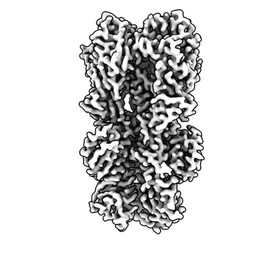 emd_40557.png emd_40557.png | 113.8 KB | ||
| Others | emd_40557_additional_1.map.gzemd_40557_additional_2.map.gzemd_40557_half_map_1.map.gzemd_40557_half_map_2.map.gz | 125.2 MB 75 MB 138.7 MB 138.7 MB | ||
| Archive directory |  http://ftp.pdbj.org/pub/emdb/structures/EMD-40557ftp://ftp.pdbj.org/pub/emdb/structures/EMD-40557 http://ftp.pdbj.org/pub/emdb/structures/EMD-40557ftp://ftp.pdbj.org/pub/emdb/structures/EMD-40557 | HTTPS FTP |
-Related structure data
| Related structure data |  8sk7MC M: atomic model generated by this map C: citing same article ( |
|---|---|
| Similar structure data |
-Links
| EMDB pages | EMDB (EBI/PDBe) / EMDataResource |
|---|---|
| Related items in Molecule of the Month |


-Map
| File | Download / File: emd_40557.map.gz / Format: CCP4 / Size: 149.9 MB / Type: IMAGE STORED AS FLOATING POINT NUMBER (4 BYTES) | ||||||||||||||||||||
|---|---|---|---|---|---|---|---|---|---|---|---|---|---|---|---|---|---|---|---|---|---|
| Annotation | unsharpened | ||||||||||||||||||||
| Voxel size | X=Y=Z: 0.83 Å | ||||||||||||||||||||
| Density |
| ||||||||||||||||||||
| Symmetry | Space group: 1 | ||||||||||||||||||||
| Details | EMDB XML:
|
-Supplemental data
-Additional map: deepEMhancer
| File | emd_40557_additional_1.map | ||||||||||||
|---|---|---|---|---|---|---|---|---|---|---|---|---|---|
| Annotation | deepEMhancer | ||||||||||||
| Projections & Slices |
| ||||||||||||
| Density Histograms |
 Z
Z Y
Y X
X


-Additional map: autosharpened
| File | emd_40557_additional_2.map | ||||||||||||
|---|---|---|---|---|---|---|---|---|---|---|---|---|---|
| Annotation | autosharpened | ||||||||||||
| Projections & Slices |
| ||||||||||||
| Density Histograms |


-Half map: half map a
| File | emd_40557_half_map_1.map | ||||||||||||
|---|---|---|---|---|---|---|---|---|---|---|---|---|---|
| Annotation | half map a | ||||||||||||
| Projections & Slices |
| ||||||||||||
| Density Histograms |


-Half map: half map b
| File | emd_40557_half_map_2.map | ||||||||||||
|---|---|---|---|---|---|---|---|---|---|---|---|---|---|
| Annotation | half map b | ||||||||||||
| Projections & Slices |
| ||||||||||||
| Density Histograms |


- Sample components
-Entire : Influenza HA (Iowa43) bound to RFdiffusion designed minibinder, HA_20
| Entire | Name: Influenza HA (Iowa43) bound to RFdiffusion designed minibinder, HA_20 |
|---|---|
| Components |
|
-Supramolecule #1: Influenza HA (Iowa43) bound to RFdiffusion designed minibinder, HA_20
| Supramolecule | Name: Influenza HA (Iowa43) bound to RFdiffusion designed minibinder, HA_20 type: complex / ID: 1 / Parent: 0 / Macromolecule list: #1-#3 |
|---|---|
| Source (natural) | Organism: Influenza A virus |
-Macromolecule #1: Hemagglutinin HA1 chain
| Macromolecule | Name: Hemagglutinin HA1 chain / type: protein_or_peptide / ID: 1 / Number of copies: 3 / Enantiomer: LEVO |
|---|---|
| Source (natural) | Organism: Influenza A virus |
| Molecular weight | Theoretical: 35.92334 KDa |
| Recombinant expression | Organism:  Homo sapiens (human) Homo sapiens (human) |
| Sequence | String: DTICIGYHAN NSTDTVDTVL EKNVTVTHSV NLLEDSHNGK LCRLKGIAPL QLGKCNIAGW ILGNPECESL LSERSWSYIV ETPNSENGT CFPGDFIDYE ELREQLSSVS SFERFEIFSK ESSWPKHTTG GVTAACSHAG KSSFYRNLLW LTEKDGSYPN L NNSYVNKK ...String: DTICIGYHAN NSTDTVDTVL EKNVTVTHSV NLLEDSHNGK LCRLKGIAPL QLGKCNIAGW ILGNPECESL LSERSWSYIV ETPNSENGT CFPGDFIDYE ELREQLSSVS SFERFEIFSK ESSWPKHTTG GVTAACSHAG KSSFYRNLLW LTEKDGSYPN L NNSYVNKK GKEVLVLWGV HHPSNIKDQQ TLYQKENAYV SVVSSNYNRR FTPEIAERPK VRGQAGRINY YWTLLKPGDT IM FEANGNL IAPWYAFALS RGFGSGIITS NASMHECDTK CQTPQGAINS SLPFQNIHPI TIGECPKYVR STKLRMVTGL RNI P UniProtKB: Hemagglutinin |
-Macromolecule #2: Hemagglutinin
| Macromolecule | Name: Hemagglutinin / type: protein_or_peptide / ID: 2 / Number of copies: 3 / Enantiomer: LEVO |
|---|---|
| Source (natural) | Organism: Influenza A virus |
| Molecular weight | Theoretical: 26.623463 KDa |
| Recombinant expression | Organism: Homo sapiens (human) |
| Sequence | String: SIQSRGLFGA IAGFIEGGWT GMIDGWYGYH WQNEQGSGYA ADQKSTQNAI NGITNIVNSV IEKMNTQFTA VGKEFNNLEK RMENLNKKV DDGFLDIWTY NAELLVLLIN ERTLDFHDSN VKNLYEKVKN QLRNNAKEIG NGCFEFYHKC NNECMESVKN G TYDYPKYS ...String: SIQSRGLFGA IAGFIEGGWT GMIDGWYGYH WQNEQGSGYA ADQKSTQNAI NGITNIVNSV IEKMNTQFTA VGKEFNNLEK RMENLNKKV DDGFLDIWTY NAELLVLLIN ERTLDFHDSN VKNLYEKVKN QLRNNAKEIG NGCFEFYHKC NNECMESVKN G TYDYPKYS EESKLNREKI DGSGYIPEAP RDGQAYVRKD GEWVLLSTFL GSGLNDIFEA QKIEWHEGHH HHHH UniProtKB: Hemagglutinin |
-Macromolecule #3: HA_20 minibinder (RFdiffusion-designed)
| Macromolecule | Name: HA_20 minibinder (RFdiffusion-designed) / type: protein_or_peptide / ID: 3 / Number of copies: 3 / Enantiomer: LEVO |
|---|---|
| Source (natural) | Organism: unidentified (others) |
| Molecular weight | Theoretical: 7.391848 KDa |
| Recombinant expression | Organism:  Escherichia coli (E. coli) Escherichia coli (E. coli) |
| Sequence | String: MEKEKELKEY AEKIKKEIGD IESVEVKDGK ILVKAKKITD KTVDAIMKLT VKAARLGFKV EVELV |
-Macromolecule #6: 2-acetamido-2-deoxy-beta-D-glucopyranose
| Macromolecule | Name: 2-acetamido-2-deoxy-beta-D-glucopyranose / type: ligand / ID: 6 / Number of copies: 12 / Formula: NAG |
|---|---|
| Molecular weight | Theoretical: 221.208 Da |
| Chemical component information |  ChemComp-NAG: |
-Experimental details
-Structure determination
| Method | cryo EM |
|---|---|
 Processing Processing | single particle reconstruction |
| Aggregation state | particle |
-Sample preparation
| Buffer | pH: 7.5 |
|---|---|
| Vitrification | Cryogen name: ETHANE |
- Electron microscopy
| Microscope | TFS KRIOS |
|---|---|
| Electron beam | Acceleration voltage: 300 kV / Electron source: FIELD EMISSION GUN |
| Electron optics | Illumination mode: FLOOD BEAM / Imaging mode: BRIGHT FIELDBright-field microscopy / Nominal defocus max: 1.7 µm / Nominal defocus min: 0.8 µm |
| Image recording | Film or detector model: GATAN K3 BIOQUANTUM (6k x 4k) / Average electron dose: 64.273 e/Å2 |
| Experimental equipment |  Model: Titan Krios / Image courtesy: FEI Company |
-Image processing
| Startup model | Type of model: PDB ENTRY PDB model - PDB ID: |
|---|---|
| Initial angle assignment | Type: MAXIMUM LIKELIHOOD |
| Final angle assignment | Type: MAXIMUM LIKELIHOOD |
| Final reconstruction | Resolution.type: BY AUTHOR / Resolution: 2.93 Å / Resolution method: FSC 0.143 CUT-OFF / Number images used: 308846 |
| FSC plot (resolution estimation) |  |