 Movie
Movie Controller


+ Open data

- Basic information
| Entry | 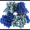 | |||||||||
|---|---|---|---|---|---|---|---|---|---|---|
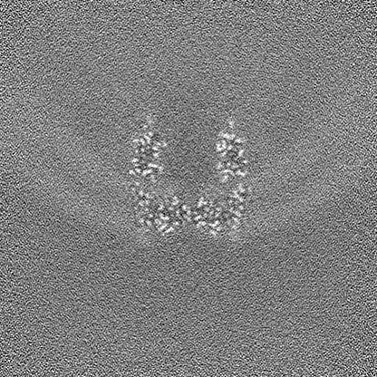
| Title | cryo-EM structure of hSlo1 in plasma membrane vesicles | |||||||||
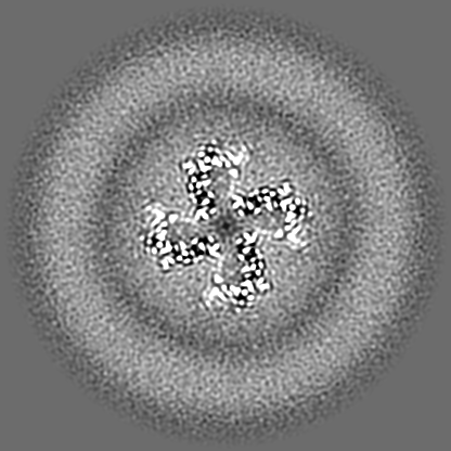
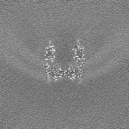
 Map data Map data | cryo-EM map of hSlo1 in plasma membrane vesicles, sharpened with a B-factor of 59.1 | |||||||||
 Sample Sample |
| |||||||||
| Function / homology |  Function and homology information Function and homology informationAcetylcholine inhibits contraction of outer hair cells / large conductance calcium-activated potassium channel activity /  micturition / Ca2+ activated K+ channels / response to carbon monoxide / calcium-activated potassium channel activity / negative regulation of cell volume / smooth muscle contraction involved in micturition / intracellular potassium ion homeostasis / Sensory processing of sound by inner hair cells of the cochlea ...Acetylcholine inhibits contraction of outer hair cells / large conductance calcium-activated potassium channel activity / micturition / Ca2+ activated K+ channels / response to carbon monoxide / calcium-activated potassium channel activity / negative regulation of cell volume / smooth muscle contraction involved in micturition / intracellular potassium ion homeostasis / Sensory processing of sound by inner hair cells of the cochlea / response to osmotic stress / cGMP effects / voltage-gated potassium channel activity / voltage-gated potassium channel complex / potassium ion transmembrane transport / regulation of membrane potential / caveola / potassium ion transport / response to calcium ion / vasodilation / actin binding / postsynaptic membrane / response to hypoxia / positive regulation of apoptotic process / apical plasma membrane / membrane / identical protein binding / metal ion binding / plasma membrane micturition / Ca2+ activated K+ channels / response to carbon monoxide / calcium-activated potassium channel activity / negative regulation of cell volume / smooth muscle contraction involved in micturition / intracellular potassium ion homeostasis / Sensory processing of sound by inner hair cells of the cochlea ...Acetylcholine inhibits contraction of outer hair cells / large conductance calcium-activated potassium channel activity / micturition / Ca2+ activated K+ channels / response to carbon monoxide / calcium-activated potassium channel activity / negative regulation of cell volume / smooth muscle contraction involved in micturition / intracellular potassium ion homeostasis / Sensory processing of sound by inner hair cells of the cochlea / response to osmotic stress / cGMP effects / voltage-gated potassium channel activity / voltage-gated potassium channel complex / potassium ion transmembrane transport / regulation of membrane potential / caveola / potassium ion transport / response to calcium ion / vasodilation / actin binding / postsynaptic membrane / response to hypoxia / positive regulation of apoptotic process / apical plasma membrane / membrane / identical protein binding / metal ion binding / plasma membraneSimilarity search - Function | |||||||||
| Biological species |  Homo sapiens (human) Homo sapiens (human) | |||||||||
| Method | single particle reconstruction / cryo EM / Resolution: 2.7 Å | |||||||||
 Authors Authors | Tao X / Zhao C / MacKinnon R | |||||||||
| Funding support |  United States, 2 items United States, 2 items
| |||||||||
 Citation Citation | Journal: Proc Natl Acad Sci U S A / Year: 2023 Title: Membrane protein isolation and structure determination in cell-derived membrane vesicles. Authors: Xiao Tao / Chen Zhao / Roderick MacKinnon / Abstract: Integral membrane protein structure determination traditionally requires extraction from cell membranes using detergents or polymers. Here, we describe the isolation and structure determination of ...Integral membrane protein structure determination traditionally requires extraction from cell membranes using detergents or polymers. Here, we describe the isolation and structure determination of proteins in membrane vesicles derived directly from cells. Structures of the ion channel Slo1 from total cell membranes and from cell plasma membranes were determined at 3.8 Å and 2.7 Å resolution, respectively. The plasma membrane environment stabilizes Slo1, revealing an alteration of global helical packing, polar lipid, and cholesterol interactions that stabilize previously unresolved regions of the channel and an additional ion binding site in the Ca regulatory domain. The two methods presented enable structural analysis of both internal and plasma membrane proteins without disrupting weakly interacting proteins, lipids, and cofactors that are essential to biological function. | |||||||||
| History |
|
- Structure visualization
| Supplemental images |
|---|

- Downloads & links
-EMDB archive
| Map data | emd_40044.map.gz | 258.9 MB | EMDB map data format | |
|---|---|---|---|---|
| Header (meta data) | emd-40044-v30.xmlemd-40044.xml | 20.5 KB 20.5 KB | Display Display | EMDB header |
| FSC (resolution estimation) | emd_40044_fsc.xml | 13.7 KB | Display | FSC data file |
| Images |  emd_40044.png emd_40044.png | 219.9 KB | ||
| Others | emd_40044_additional_1.map.gzemd_40044_half_map_1.map.gzemd_40044_half_map_2.map.gz | 133.7 MB 254.5 MB 254.5 MB | ||
| Archive directory |  http://ftp.pdbj.org/pub/emdb/structures/EMD-40044ftp://ftp.pdbj.org/pub/emdb/structures/EMD-40044 http://ftp.pdbj.org/pub/emdb/structures/EMD-40044ftp://ftp.pdbj.org/pub/emdb/structures/EMD-40044 | HTTPS FTP |
-Related structure data
| Related structure data |  8ghfMC  8gh9C 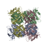 8ghgC M: atomic model generated by this map C: citing same article ( |
|---|---|
| Similar structure data |

-Links
| EMDB pages | EMDB (EBI/PDBe) / EMDataResource |
|---|---|
| Related items in Molecule of the Month |


-Map
| File | Download / File: emd_40044.map.gz / Format: CCP4 / Size: 274.6 MB / Type: IMAGE STORED AS FLOATING POINT NUMBER (4 BYTES) | ||||||||||||||||||||
|---|---|---|---|---|---|---|---|---|---|---|---|---|---|---|---|---|---|---|---|---|---|
| Annotation | cryo-EM map of hSlo1 in plasma membrane vesicles, sharpened with a B-factor of 59.1 | ||||||||||||||||||||
| Voxel size | X=Y=Z: 0.743 Å | ||||||||||||||||||||
| Density |
| ||||||||||||||||||||
| Symmetry | Space group: 1 | ||||||||||||||||||||
| Details | EMDB XML:
|
-Supplemental data
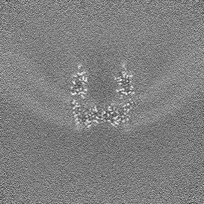
-Additional map: unsharpened cryo-EM map of hSlo1 in plasma membrane vesicles
| File | emd_40044_additional_1.map | ||||||||||||
|---|---|---|---|---|---|---|---|---|---|---|---|---|---|
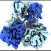
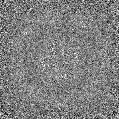
| Annotation | unsharpened cryo-EM map of hSlo1 in plasma membrane vesicles | ||||||||||||
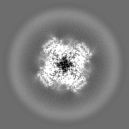
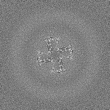
| Projections & Slices |
| ||||||||||||
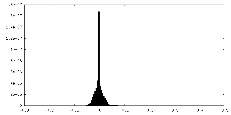
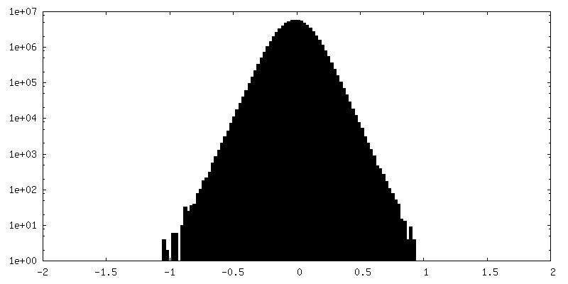
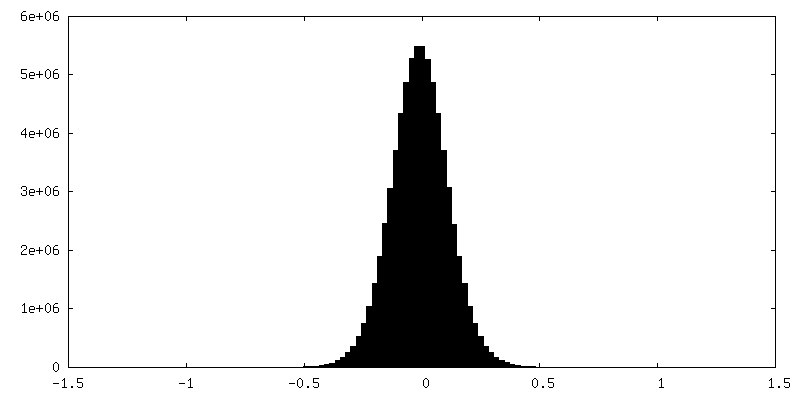
| Density Histograms |
 Z
Z Y
Y X
X


-Half map: half map 1 of hSlo1 in plasma membrane vesicles
| File | emd_40044_half_map_1.map | ||||||||||||
|---|---|---|---|---|---|---|---|---|---|---|---|---|---|
| Annotation | half map 1 of hSlo1 in plasma membrane vesicles | ||||||||||||
| Projections & Slices |
| ||||||||||||
| Density Histograms |


-Half map: half map 2 of hSlo1 in plasma membrane vesicles
| File | emd_40044_half_map_2.map | ||||||||||||
|---|---|---|---|---|---|---|---|---|---|---|---|---|---|
| Annotation | half map 2 of hSlo1 in plasma membrane vesicles | ||||||||||||
| Projections & Slices |
| ||||||||||||
| Density Histograms |


- Sample components
-Entire : ALFA-hSlo1-eGFP ion channel
| Entire | Name: ALFA-hSlo1-eGFP ion channel |
|---|---|
| Components |
|
-Supramolecule #1: ALFA-hSlo1-eGFP ion channel
| Supramolecule | Name: ALFA-hSlo1-eGFP ion channel / type: complex / ID: 1 / Chimera: Yes / Parent: 0 / Macromolecule list: #1 |
|---|---|
| Source (natural) | Organism: Homo sapiens (human) |
-Macromolecule #1: Calcium-activated potassium channel subunit alpha-1
| Macromolecule | Name: Calcium-activated potassium channel subunit alpha-1 / type: protein_or_peptide / ID: 1 / Number of copies: 4 / Enantiomer: LEVO |
|---|---|
| Source (natural) | Organism: Homo sapiens (human) |
| Molecular weight | Theoretical: 122.440102 KDa |
| Recombinant expression | Organism: Homo sapiens (human) |
| Sequence | String: MAPSRLEEEL RRRLTEPDAL IIPVTMEVPC DSRGQRMWWA FLASSMVTFF GGLFIILLWR TLKYLWTVCC HCGGKTKEAQ KINNGSSQA DGTLKPVDEK EEAVAAEVGW MTSVKDWAGV MISAQTLTGR VLVVLVFALS IGALVIYFID SSNPIESCQN F YKDFTLQI ...String: MAPSRLEEEL RRRLTEPDAL IIPVTMEVPC DSRGQRMWWA FLASSMVTFF GGLFIILLWR TLKYLWTVCC HCGGKTKEAQ KINNGSSQA DGTLKPVDEK EEAVAAEVGW MTSVKDWAGV MISAQTLTGR VLVVLVFALS IGALVIYFID SSNPIESCQN F YKDFTLQI DMAFNVFFLL YFGLRFIAAN DKLWFWLEVN SVVDFFTVPP VFVSVYLNRS WLGLRFLRAL RLIQFSEILQ FL NILKTSN SIKLVNLLSI FISTWLTAAG FIHLVENSGD PWENFQNNQA LTYWECVYLL MVTMSTVGYG DVYAKTTLGR LFM VFFILG GLAMFASYVP EIIELIGNRK KYGGSYSAVS GRKHIVVCGH ITLESVSNFL KDFLHKDRDD VNVEIVFLHN ISPN LELEA LFKRHFTQVE FYQGSVLNPH DLARVKIESA DACLILANKY CADPDAEDAS NIMRVISIKN YHPKIRIITQ MLQYH NKAH LLNIPSWNWK EGDDAICLAE LKLGFIAQSC LAQGLSTMLA NLFSMRSFIK IEEDTWQKYY LEGVSNEMYT EYLSSA FVG LSFPTVCELC FVKLKLLMIA IEYKSANRES RILINPGNHL KIQEGTLGFF IASDAKEVKR AFFYCKACHD DITDPKR IK KCGCKRLEDE QPSTLSPKKK QRNGGMRNSP NTSPKLMRHD PLLIPGNDQI DNMDSNVKKY DSTGMFHWCA PKEIEKVI L TRSEAAMTVL SGHVVVCIFG DVSSALIGLR NLVMPLRASN FHYHELKHIV FVGSIEYLKR EWETLHNFPK VSILPGTPL SRADLRAVNI NLCDMCVILS ANQNNIDDTS LQDKECILAS LNIKSMQFDD SIGVLQANSQ GFTPPGMDRS SPDNSPVHGM LRQPSITTG VNIPIITELV NDTNVQFLDQ DDDDDPDTEL YLTQPFACGT AFAVSVLDSL MSATYFNDNI LTLIRTLVTG G ATPELEAL IAEENALRGG YSTPQTLANR DRCRVAQLAL LDGPFADLGD GGCYGDLFCK ALKTYNMLCF GIYRLRDAHL ST PSQCTKR YVITNPPYEF ELVPTDLIFC LMQFD(UNK)(UNK)(UNK)(UNK)(UNK) (UNK)(UNK)(UNK)(UNK) (UNK)(UNK)(UNK)(UNK)(UNK) (UNK)(UNK)(UNK)(UNK) |
-Macromolecule #2: MAGNESIUM ION
| Macromolecule | Name: MAGNESIUM ION / type: ligand / ID: 2 / Number of copies: 4 / Formula: MG |
|---|---|
| Molecular weight | Theoretical: 24.305 Da |
-Macromolecule #3: CALCIUM ION
| Macromolecule | Name: CALCIUM ION / type: ligand / ID: 3 / Number of copies: 8 / Formula: CA |
|---|---|
| Molecular weight | Theoretical: 40.078 Da |
-Macromolecule #4: (2S)-3-(hexadecanoyloxy)-2-[(9Z)-octadec-9-enoyloxy]propyl 2-(tri...
| Macromolecule | Name: (2S)-3-(hexadecanoyloxy)-2-[(9Z)-octadec-9-enoyloxy]propyl 2-(trimethylammonio)ethyl phosphate type: ligand / ID: 4 / Number of copies: 88 / Formula: POV |
|---|---|
| Molecular weight | Theoretical: 760.076 Da |
| Chemical component information |  ChemComp-POV: |
-Macromolecule #5: CHOLESTEROL
| Macromolecule | Name: CHOLESTEROL / type: ligand / ID: 5 / Number of copies: 24 / Formula: CLR |
|---|---|
| Molecular weight | Theoretical: 386.654 Da |
| Chemical component information |  ChemComp-CLR: |
-Macromolecule #6: SODIUM ION
| Macromolecule | Name: SODIUM ION / type: ligand / ID: 6 / Number of copies: 4 |
|---|---|
| Molecular weight | Theoretical: 22.99 Da |
-Experimental details
-Structure determination
| Method | cryo EM |
|---|---|
 Processing Processing | single particle reconstruction |
| Aggregation state | particle |
-Sample preparation
| Buffer | pH: 7.4 |
|---|---|
| Grid | Model: Quantifoil R1.2/1.3 / Material: GOLD / Mesh: 400 / Pretreatment - Type: GLOW DISCHARGE / Pretreatment - Time: 22 sec. |
| Vitrification | Cryogen name: ETHANE / Chamber humidity: 100 % / Chamber temperature: 295.15 K / Instrument: FEI VITROBOT MARK IV |
- Electron microscopy
| Microscope | FEI TITAN KRIOS |
|---|---|
| Electron beam | Acceleration voltage: 300 kV / Electron source: FIELD EMISSION GUN |
| Electron optics | Illumination mode: FLOOD BEAM / Imaging mode: BRIGHT FIELDBright-field microscopy / Cs: 2.7 mm / Nominal defocus max: 2.0 µm / Nominal defocus min: 1.0 µm |
| Specialist optics | Energy filter - Slit width: 6 eV |
| Sample stage | Specimen holder model: FEI TITAN KRIOS AUTOGRID HOLDER / Cooling holder cryogen: NITROGEN |
| Image recording | Film or detector model: FEI FALCON IV (4k x 4k) / Average electron dose: 60.0 e/Å2 |
| Experimental equipment |  Model: Titan Krios / Image courtesy: FEI Company |
-Image processing
| Startup model | Type of model: OTHER / Details: ab-initio model by cryoSPARC |
|---|---|
| Initial angle assignment | Type: MAXIMUM LIKELIHOOD / Software - Name: cryoSPARC |
| Final 3D classification | Software - Name: RELION |
| Final angle assignment | Type: MAXIMUM LIKELIHOOD / Software - Name: cryoSPARC |
| Final reconstruction | Applied symmetry - Point group: C4 (4 fold cyclic) / Algorithm: BACK PROJECTION / Resolution.type: BY AUTHOR / Resolution: 2.7 Å / Resolution method: FSC 0.143 CUT-OFF / Software - Name: cryoSPARC / Number images used: 32063 |
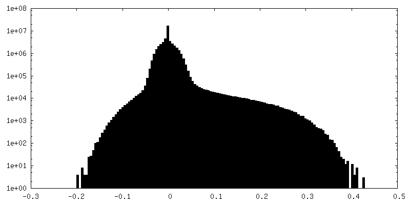
| FSC plot (resolution estimation) |  |
