organelle inner membrane / plasma membrane-derived chromatophore membrane / plasma membrane light-harvesting complex / bacteriochlorophyll binding / photosynthesis, light reaction / electron transporter, transferring electrons within the cyclic electron transport pathway of photosynthesis activity / photosynthetic electron transport in photosystem II / electron transfer activity / iron ion binding / heme binding ...organelle inner membrane / plasma membrane-derived chromatophore membrane / plasma membrane light-harvesting complex / bacteriochlorophyll binding / photosynthesis, light reaction / electron transporter, transferring electrons within the cyclic electron transport pathway of photosynthesis activity / photosynthetic electron transport in photosystem II / electron transfer activity / iron ion binding / heme binding / membrane / metal ion binding / plasma membrane Similarity search - Function
Photosynthetic reaction centre, cytochrome c subunit / Multihaem cytochrome, PRC subunit superfamily / Photosynthetic reaction centre cytochrome C subunit / Antenna complex, beta subunit, conserved site / Antenna complexes beta subunits signature. / Antenna complex, alpha subunit / Antenna complex, beta domain superfamily / Antenna complex, alpha subunit conserved site / Antenna complexes alpha subunits signature. / Light-harvesting protein B beta chain ...Photosynthetic reaction centre, cytochrome c subunit / Multihaem cytochrome, PRC subunit superfamily / Photosynthetic reaction centre cytochrome C subunit / Antenna complex, beta subunit, conserved site / Antenna complexes beta subunits signature. / Antenna complex, alpha subunit / Antenna complex, beta domain superfamily / Antenna complex, alpha subunit conserved site / Antenna complexes alpha subunits signature. / Light-harvesting protein B beta chain / Antenna complex, alpha/beta subunit / Light-harvesting complex / Antenna complex alpha/beta subunit / Photosynthetic reaction centre, H subunit / Bacterial photosynthetic reaction centre, H-chain, C-terminal / Photosynthetic reaction centre, M subunit / Photosynthetic reaction centre, H subunit, N-terminal / Photosynthetic reaction centre, H subunit, N-terminal domain superfamily / Photosynthetic reaction centre, H-chain N-terminal region / PRC-barrel domain / PRC-barrel domain / Photosynthetic reaction centre, L subunit / PRC-barrel-like superfamily / Multiheme cytochrome superfamily / Photosynthetic reaction centre, L/M / Photosystem II protein D1/D2 superfamily / Photosynthetic reaction centre protein / Photosynthetic reaction center proteins signature. / Prokaryotic membrane lipoprotein lipid attachment site profile. Similarity search - Domain/homology
Photosynthetic reaction center cytochrome c subunit / Photosynthetic reaction center H subunit / Reaction center protein M chain / Uncharacterized protein / Reaction center protein L chain / Antenna complex alpha/beta subunit domain-containing protein / Antenna complex alpha/beta subunit domain-containing protein / Light-harvesting LHI Similarity search - Component
Biological species
Halorhodospira halochloris (bacteria)
Method
single particle reconstruction / cryo EM / Resolution: 2.42 Å
Journal: J Integr Plant Biol / Year: 2024 Title: Structural insights into the unusual core photocomplex from a triply extremophilic purple bacterium, Halorhodospira halochloris. Authors: Chen-Hui Qi / Guang-Lei Wang / Fang-Fang Wang / Jie Wang / Xiang-Ping Wang / Mei-Juan Zou / Fei Ma / Michael T Madigan / Yukihiro Kimura / Zheng-Yu Wang-Otomo / Long-Jiang Yu / Abstract: Halorhodospira (Hlr.) halochloris is a triply extremophilic phototrophic purple sulfur bacterium, as it is thermophilic, alkaliphilic, and extremely halophilic. The light-harvesting-reaction center ...Halorhodospira (Hlr.) halochloris is a triply extremophilic phototrophic purple sulfur bacterium, as it is thermophilic, alkaliphilic, and extremely halophilic. The light-harvesting-reaction center (LH1-RC) core complex of this bacterium displays an LH1-Q transition at 1,016 nm, which is the lowest-energy wavelength absorption among all known phototrophs. Here we report the cryo-EM structure of the LH1-RC at 2.42 Å resolution. The LH1 complex forms a tricyclic ring structure composed of 16 αβγ-polypeptides and one αβ-heterodimer around the RC. From the cryo-EM density map, two previously unrecognized integral membrane proteins, referred to as protein G and protein Q, were identified. Both of these proteins are single transmembrane-spanning helices located between the LH1 ring and the RC L-subunit and are absent from the LH1-RC complexes of all other purple bacteria of which the structures have been determined so far. Besides bacteriochlorophyll b molecules (B1020) located on the periplasmic side of the Hlr. halochloris membrane, there are also two arrays of bacteriochlorophyll b molecules (B800 and B820) located on the cytoplasmic side. Only a single copy of a carotenoid (lycopene) was resolved in the Hlr. halochloris LH1-α3β3 and this was positioned within the complex. The potential quinone channel should be the space between the LH1-α3β3 that accommodates the single lycopene but does not contain a γ-polypeptide, B800 and B820. Our results provide a structural explanation for the unusual Q red shift and carotenoid absorption in the Hlr. halochloris spectrum and reveal new insights into photosynthetic mechanisms employed by a species that thrives under the harshest conditions of any phototrophic microorganism known.
In the structure databanks used in Yorodumi, some data are registered as the other names, "COVID-19 virus" and "2019-nCoV". Here are the details of the virus and the list of structure data.
Jan 31, 2019. EMDB accession codes are about to change! (news from PDBe EMDB page)
EMDB accession codes are about to change! (news from PDBe EMDB page)
The allocation of 4 digits for EMDB accession codes will soon come to an end. Whilst these codes will remain in use, new EMDB accession codes will include an additional digit and will expand incrementally as the available range of codes is exhausted. The current 4-digit format prefixed with “EMD-” (i.e. EMD-XXXX) will advance to a 5-digit format (i.e. EMD-XXXXX), and so on. It is currently estimated that the 4-digit codes will be depleted around Spring 2019, at which point the 5-digit format will come into force.
The EM Navigator/Yorodumi systems omit the EMD- prefix.
Related info.:Q: What is EMD? / ID/Accession-code notation in Yorodumi/EM Navigator
Yorodumi is a browser for structure data from EMDB, PDB, SASBDB, etc.
This page is also the successor to EM Navigator detail page, and also detail information page/front-end page for Omokage search.
The word "yorodu" (or yorozu) is an old Japanese word meaning "ten thousand". "mi" (miru) is to see.
Related info.:EMDB / PDB / SASBDB / Comparison of 3 databanks / Yorodumi Search / Aug 31, 2016. New EM Navigator & Yorodumi / Yorodumi Papers / Jmol/JSmol / Function and homology information / Changes in new EM Navigator and Yorodumi
 Movie
Movie Controller

 Yorodumi
 Open data
 Basic information
 Map data
 Sample
 Keywords
 PHOTOSYNTHESIS
 Function and homology information
 Halorhodospira halochloris (bacteria)
 Authors
 China, 1 items
  Citation

 Structure visualization
 Downloads & links
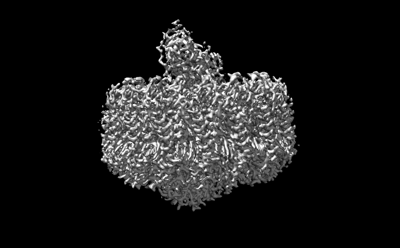 emd_36907.png
 http://ftp.pdbj.org/pub/emdb/structures/EMD-36907


 Z
Z Y
Y X
X


 Sample components


 Processing
 Electron microscopy

