 Movie
Movie Controller


+ Open data

- Basic information
| Entry |  | |||||||||
|---|---|---|---|---|---|---|---|---|---|---|
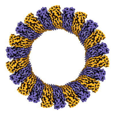
| Title | Cryo-EM structure of RCD-1 pore from Neurospora crassa | |||||||||
 Map data Map data | ||||||||||
 Sample Sample |
| |||||||||
 Keywords Keywords |  Pyroptosis / Gasdermnin / Pore / Allorecognition / IMMUNE SYSTEM Pyroptosis / Gasdermnin / Pore / Allorecognition / IMMUNE SYSTEM | |||||||||
| Function / homology | wide pore channel activity / programmed cell death / protein heterodimerization activity / plasma membrane / cytoplasm / Gasdermin-like protein rcd-1-2 / Gasdermin-like protein rcd-1-1 Function and homology information Function and homology information | |||||||||
| Biological species |  Neurospora crassa (fungus) Neurospora crassa (fungus) | |||||||||
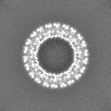
| Method | single particle reconstruction / cryo EM / Resolution: 3.63 Å | |||||||||
 Authors Authors | Hou YJ / Sun Q / Li Y / Ding J | |||||||||
| Funding support |  China, 1 items China, 1 items
| |||||||||
 Citation Citation | Journal: Science / Year: 2024 Title: Cleavage-independent activation of ancient eukaryotic gasdermins and structural mechanisms. Authors: Yueyue Li / Yanjie Hou / Qi Sun / Huan Zeng / Fanyi Meng / Xiang Tian / Qun He / Feng Shao / Jingjin Ding / Abstract: Gasdermins (GSDMs) are pore-forming proteins that execute pyroptosis for immune defense. GSDMs are two-domain proteins, activated by proteolytic removal of the inhibitory domain. Here we report two ...Gasdermins (GSDMs) are pore-forming proteins that execute pyroptosis for immune defense. GSDMs are two-domain proteins, activated by proteolytic removal of the inhibitory domain. Here we report two types of cleavage-independent GSDM activation. First, GSDM, a pore-forming-domain-only protein from the basal metazoan , is a disulfides-linked autoinhibited dimer, activated by reduction of the disulfides. Cryo-electron microscopy (cryo-EM) structure illustrates assembly mechanism for the 44-mer GSDM pore. Second, RCD-1-1/RCD-1-2, encoded by polymorphic in filamentous fungus , are also pore-forming-domain-only GSDMs. RCD-1-1 and RCD-1-2, when encountering each other, form pores and cause pyroptosis, underlying allorecognition in . Cryo-EM structure reveals a pore of 11 RCD-1-1/RCD-1-2 heterodimers and heterodimerization-triggered pore assembly mechanism. This study shows mechanistic diversities in GSDM activation and indicates versatile functions of GSDMs. | |||||||||
| History |
|
- Structure visualization
| Supplemental images |
|---|
- Downloads & links
-EMDB archive
| Map data | emd_36734.map.gz | 197.4 MB | EMDB map data format | |
|---|---|---|---|---|
| Header (meta data) | emd-36734-v30.xmlemd-36734.xml | 17 KB 17 KB | Display Display | EMDB header |
| FSC (resolution estimation) | emd_36734_fsc.xml | 13.2 KB | Display | FSC data file |
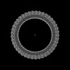
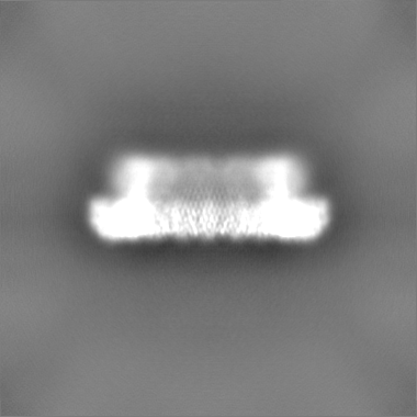
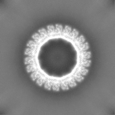
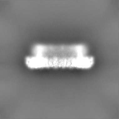
| Images |  emd_36734.png emd_36734.png | 178.8 KB | ||
| Filedesc metadata | emd-36734.cif.gz | 5.8 KB | ||
| Others | emd_36734_half_map_1.map.gzemd_36734_half_map_2.map.gz | 193.5 MB 193.6 MB | ||
| Archive directory |  http://ftp.pdbj.org/pub/emdb/structures/EMD-36734ftp://ftp.pdbj.org/pub/emdb/structures/EMD-36734 http://ftp.pdbj.org/pub/emdb/structures/EMD-36734ftp://ftp.pdbj.org/pub/emdb/structures/EMD-36734 | HTTPS FTP |
-Related structure data
| Related structure data |  8jyzMC 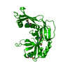 8jyvC 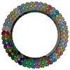 8jywC 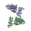 8jyxC 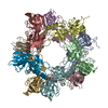 8jyyC M: atomic model generated by this map C: citing same article ( |
|---|---|
| Similar structure data |

-Links
| EMDB pages | EMDB (EBI/PDBe) / EMDataResource |
|---|
-Map
| File | Download / File: emd_36734.map.gz / Format: CCP4 / Size: 209.3 MB / Type: IMAGE STORED AS FLOATING POINT NUMBER (4 BYTES) | ||||||||||||||||||||
|---|---|---|---|---|---|---|---|---|---|---|---|---|---|---|---|---|---|---|---|---|---|
| Voxel size | X=Y=Z: 1.07 Å | ||||||||||||||||||||
| Density |
| ||||||||||||||||||||
| Symmetry | Space group: 1 | ||||||||||||||||||||
| Details | EMDB XML:
|
-Supplemental data
-Half map: #1
| File | emd_36734_half_map_1.map | ||||||||||||
|---|---|---|---|---|---|---|---|---|---|---|---|---|---|
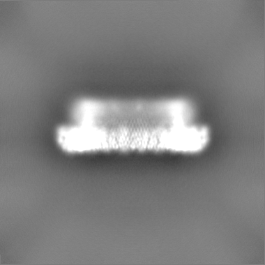
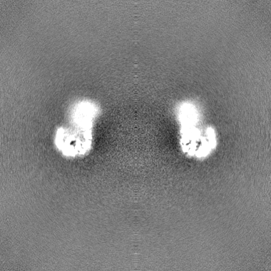
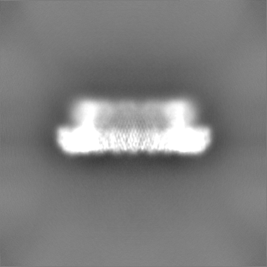
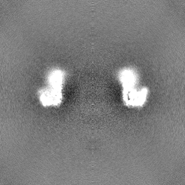
| Projections & Slices |
| ||||||||||||
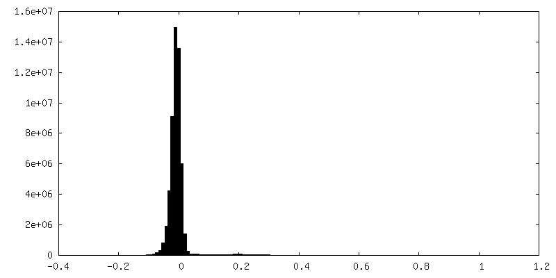
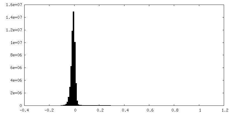
| Density Histograms |
 Z
Z Y
Y X
X


-Half map: #2
| File | emd_36734_half_map_2.map | ||||||||||||
|---|---|---|---|---|---|---|---|---|---|---|---|---|---|
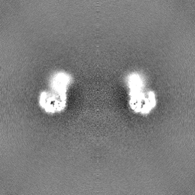
| Projections & Slices |
| ||||||||||||
| Density Histograms |

- Sample components
-Entire : RCD-1 pore from Neurospora crassa
| Entire | Name: RCD-1 pore from Neurospora crassa |
|---|---|
| Components |
|
-Supramolecule #1: RCD-1 pore from Neurospora crassa
| Supramolecule | Name: RCD-1 pore from Neurospora crassa / type: complex / ID: 1 / Parent: 0 / Macromolecule list: all |
|---|---|
| Source (natural) | Organism: Neurospora crassa (fungus) |
| Molecular weight | Theoretical: 620 KDa |
-Macromolecule #1: Gasdermin-like protein rcd-1-1
| Macromolecule | Name: Gasdermin-like protein rcd-1-1 / type: protein_or_peptide / ID: 1 / Number of copies: 11 / Enantiomer: LEVO |
|---|---|
| Source (natural) | Organism: Neurospora crassa (fungus) |
| Molecular weight | Theoretical: 29.218693 KDa |
| Recombinant expression | Organism:  Escherichia coli BL21(DE3) (bacteria) Escherichia coli BL21(DE3) (bacteria) |
| Sequence | String: SGRPMDKCWF TLDNAHYPPP SLDSMRSGHP ISPASLGHLI PSLAHLDQII NAKAIEPFPA TMDIHGPTII EDFKWDHSHE YSLSLGGKV PIPLAPAGVP FVDLNVGLGG AFSRSVANYW EFDRLERYIM QPTRSYVQKC IERDEVKRWI AKNKSMMMMG R WEVYMITG ...String: SGRPMDKCWF TLDNAHYPPP SLDSMRSGHP ISPASLGHLI PSLAHLDQII NAKAIEPFPA TMDIHGPTII EDFKWDHSHE YSLSLGGKV PIPLAPAGVP FVDLNVGLGG AFSRSVANYW EFDRLERYIM QPTRSYVQKC IERDEVKRWI AKNKSMMMMG R WEVYMITG IIVARGGGRK KKEKTTGKEF SVEVTVEVPL IVEAGPGGKR NTARQKTWGT SQTGDFVWAV RLAKITKSGL HS DWKMETV FGKTSSFRGQ KAIF UniProtKB: Gasdermin-like protein rcd-1-1 |
-Macromolecule #2: Gasdermin-like protein rcd-1-2
| Macromolecule | Name: Gasdermin-like protein rcd-1-2 / type: protein_or_peptide / ID: 2 / Number of copies: 11 / Enantiomer: LEVO |
|---|---|
| Source (natural) | Organism: Neurospora crassa (fungus) |
| Molecular weight | Theoretical: 27.630418 KDa |
| Recombinant expression | Organism: Escherichia coli BL21(DE3) (bacteria) |
| Sequence | String: SGRPMDNEEW FPLKQTHYPP PTIPSMKTGH PTGPISIGHI IPDLRHLDNV INCKGFEPFP PNMDVFTAHY EQCHFGDHLN SEFVVQAKA AAPIENIVPG VDVTGSAGLH HTNITSDRWE YDSVVEYAVY PTRQYIDRLL ESKEVRQYIQ KSKKLLGGWC V YMVTGIMV ...String: SGRPMDNEEW FPLKQTHYPP PTIPSMKTGH PTGPISIGHI IPDLRHLDNV INCKGFEPFP PNMDVFTAHY EQCHFGDHLN SEFVVQAKA AAPIENIVPG VDVTGSAGLH HTNITSDRWE YDSVVEYAVY PTRQYIDRLL ESKEVRQYIQ KSKKLLGGWC V YMVTGIMV ARGGGRNVTS EEKGAGVSGN VGFQVPGIGE FAPEVGWDTK TKTKVNAHHT TDFVCAIRLV KIAKSGLRSS WT MKKVTRE F UniProtKB: Gasdermin-like protein rcd-1-2 |
-Experimental details
-Structure determination
| Method | cryo EM |
|---|---|
 Processing Processing | single particle reconstruction |
| Aggregation state | particle |
-Sample preparation
| Buffer | pH: 8 Component:
| ||||||||||
|---|---|---|---|---|---|---|---|---|---|---|---|
| Grid | Model: Quantifoil R1.2/1.3 / Material: GOLD / Mesh: 300 / Support film - Material: GRAPHENE OXIDE / Pretreatment - Type: GLOW DISCHARGE | ||||||||||
| Vitrification | Cryogen name: ETHANE / Chamber humidity: 100 % / Chamber temperature: 277 K |
- Electron microscopy
| Microscope | FEI TITAN KRIOS |
|---|---|
| Electron beam | Acceleration voltage: 300 kV / Electron source: FIELD EMISSION GUN |
| Electron optics | Illumination mode: FLOOD BEAM / Imaging mode: BRIGHT FIELDBright-field microscopy / Nominal defocus max: 3.0 µm / Nominal defocus min: 1.5 µm |
| Image recording | Film or detector model: GATAN K3 (6k x 4k) / Average electron dose: 56.0 e/Å2 |
| Experimental equipment |  Model: Titan Krios / Image courtesy: FEI Company |
-Image processing
| Startup model | Type of model: NONE |
|---|---|
| Initial angle assignment | Type: MAXIMUM LIKELIHOOD |
| Final angle assignment | Type: PROJECTION MATCHING |
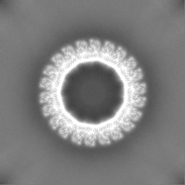
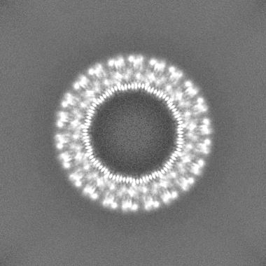
| Final reconstruction | Applied symmetry - Point group: C11 (11 fold cyclic) / Resolution.type: BY AUTHOR / Resolution: 3.63 Å / Resolution method: FSC 0.143 CUT-OFF / Number images used: 118156 |
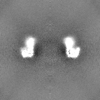
| FSC plot (resolution estimation) | 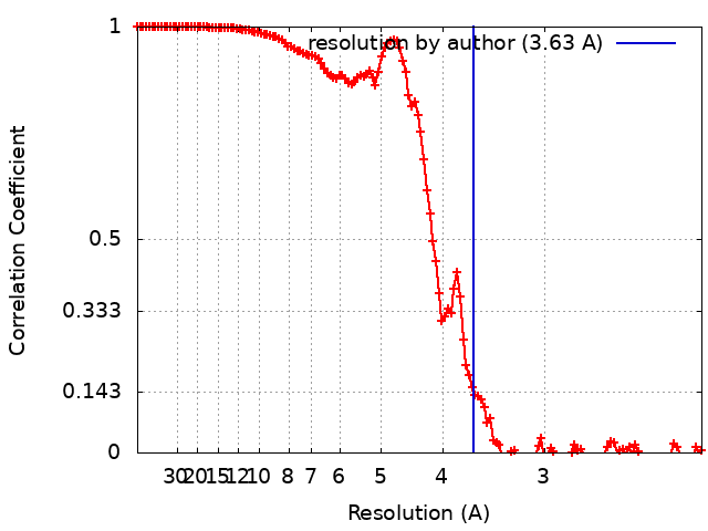 |
