Movie
Movie Controller

[English] 日本語
 Yorodumi
- EMDB-36223: Cryo-EM structure of the GI.4 Chiba VLP complexed with the CV-1A1... -
+ Open data

- Basic information
| Entry | 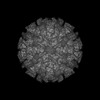 | |||||||||
|---|---|---|---|---|---|---|---|---|---|---|
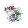
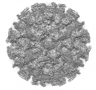
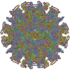
| Title | Cryo-EM structure of the GI.4 Chiba VLP complexed with the CV-1A1 Fv-clasp | |||||||||
 Map data Map data | ||||||||||
 Sample Sample |
| |||||||||
 Keywords Keywords |  NOROVIRUS / GI.4 / VIRUS LIKE PARTICLE / human monoclonal antibody / Fv-clasp / VIRUS NOROVIRUS / GI.4 / VIRUS LIKE PARTICLE / human monoclonal antibody / Fv-clasp / VIRUS | |||||||||
| Biological species |  Norovirus / Norovirus /  Homo sapiens (human) Homo sapiens (human) | |||||||||
| Method | single particle reconstruction / cryo EM / Resolution: 3.04 Å | |||||||||
 Authors Authors | Hosaka T / Katsura K / Kimura-Someya T / Someya Y / Shirouzu M | |||||||||
| Funding support |  Japan, 2 items Japan, 2 items
| |||||||||
 Citation Citation | Journal: To Be Published Title: Structural analyses of the GI.4 norovirus by cryo-electron microscopy and X-ray crystallography reveal binding sites for human monoclonal antibodies Authors: Kimura-Someya T / Katsura K / Kato-Murayama M / Hosaka T / Uchikubo-Kamo T / Ihara K / Hanada K / Murayama K / Shirouzu M / Someya Y | |||||||||
| History |
|
- Structure visualization
| Supplemental images |
|---|
- Downloads & links
-EMDB archive
| Map data | emd_36223.map.gz | 449.3 MB |  EMDB map data format EMDB map data format | |
|---|---|---|---|---|
| Header (meta data) | emd-36223-v30.xmlemd-36223.xml | 18.4 KB 18.4 KB | Display Display | EMDB header |
| FSC (resolution estimation) | emd_36223_fsc.xml | 17.5 KB | Display | FSC data file |
| Images |  emd_36223.png emd_36223.png | 191.9 KB | ||
| Masks | emd_36223_msk_1.map | 476.8 MB | Mask map | |
| Filedesc metadata | emd-36223.cif.gz | 6 KB | ||
| Others | emd_36223_additional_1.map.gzemd_36223_half_map_1.map.gzemd_36223_half_map_2.map.gz | 13.2 MB 441.2 MB 441.2 MB | ||
| Archive directory |  http://ftp.pdbj.org/pub/emdb/structures/EMD-36223ftp://ftp.pdbj.org/pub/emdb/structures/EMD-36223 http://ftp.pdbj.org/pub/emdb/structures/EMD-36223ftp://ftp.pdbj.org/pub/emdb/structures/EMD-36223 | HTTPS FTP |
-Related structure data
-Links
| EMDB pages | EMDB (EBI/PDBe) / EMDataResource |
|---|
-Map
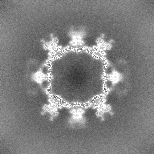
| File | Download / File: emd_36223.map.gz / Format: CCP4 / Size: 476.8 MB / Type: IMAGE STORED AS FLOATING POINT NUMBER (4 BYTES) | ||||||||||||||||||||||||||||||||||||
|---|---|---|---|---|---|---|---|---|---|---|---|---|---|---|---|---|---|---|---|---|---|---|---|---|---|---|---|---|---|---|---|---|---|---|---|---|---|
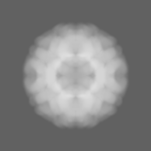
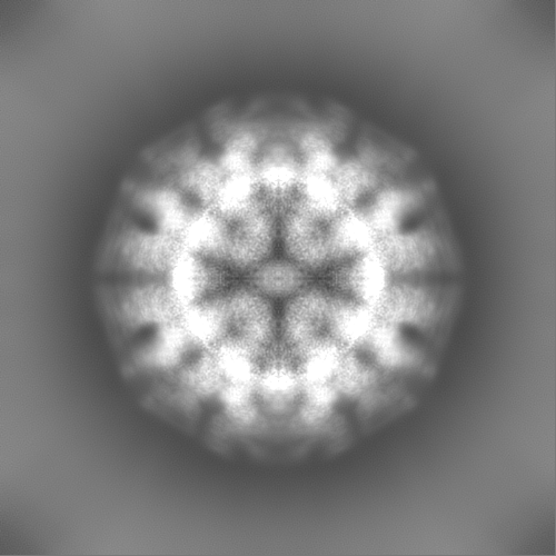
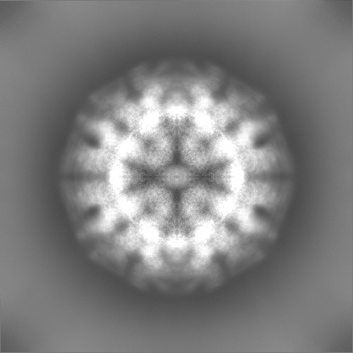
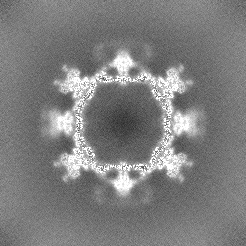
| Projections & slices | Image control
Images are generated by Spider. | ||||||||||||||||||||||||||||||||||||
| Voxel size | X=Y=Z: 1.47 Å | ||||||||||||||||||||||||||||||||||||
| Density |
| ||||||||||||||||||||||||||||||||||||
| Symmetry | Space group: 1 | ||||||||||||||||||||||||||||||||||||
| Details | EMDB XML:
|
 Z (Sec.)
 Y (Row.)
 X (Col.)


-Supplemental data
-Mask #1
| File | emd_36223_msk_1.map | ||||||||||||
|---|---|---|---|---|---|---|---|---|---|---|---|---|---|
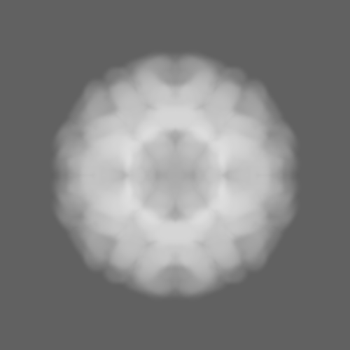
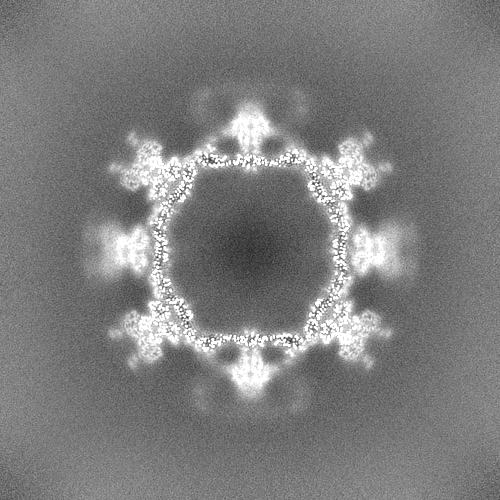
| Projections & Slices |
| ||||||||||||
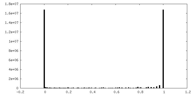
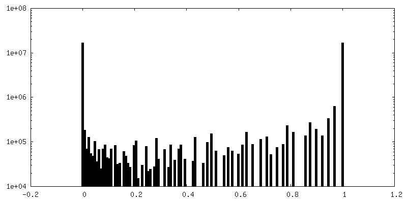
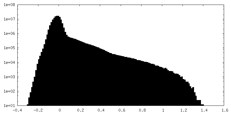
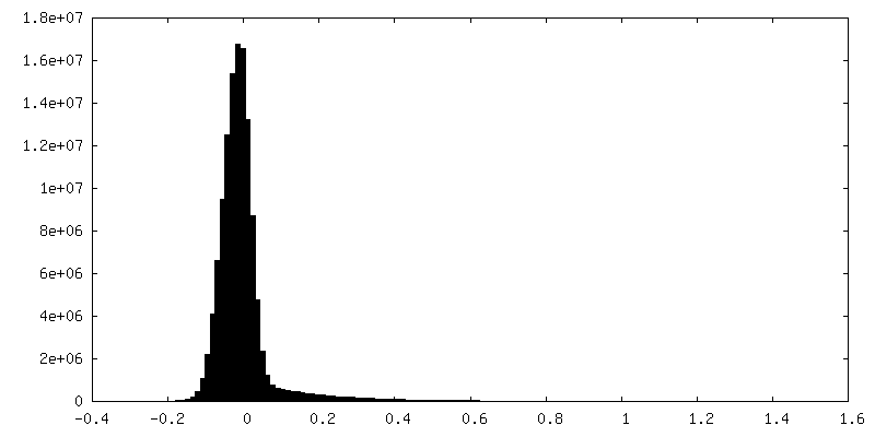
| Density Histograms |


-Additional map: #1
| File | emd_36223_additional_1.map | ||||||||||||
|---|---|---|---|---|---|---|---|---|---|---|---|---|---|
| Projections & Slices |
| ||||||||||||
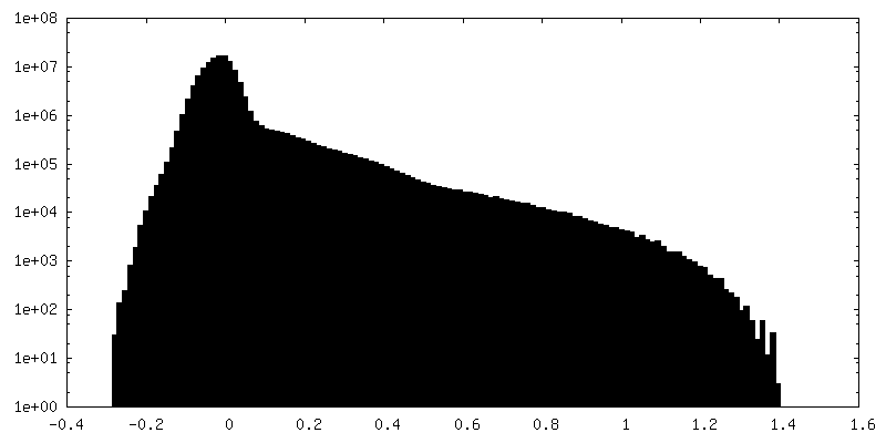
| Density Histograms |


-Half map: #1
| File | emd_36223_half_map_1.map | ||||||||||||
|---|---|---|---|---|---|---|---|---|---|---|---|---|---|
| Projections & Slices |
| ||||||||||||
| Density Histograms |


-Half map: #2
| File | emd_36223_half_map_2.map | ||||||||||||
|---|---|---|---|---|---|---|---|---|---|---|---|---|---|
| Projections & Slices |
| ||||||||||||
| Density Histograms |

- Sample components
-Entire : Viral protein 1 with its antibody fragments
| Entire | Name: Viral protein 1 with its antibody fragments |
|---|---|
| Components |
|
-Supramolecule #1: Viral protein 1 with its antibody fragments
| Supramolecule | Name: Viral protein 1 with its antibody fragments / type: complex / ID: 1 / Parent: 0 / Macromolecule list: all |
|---|---|
| Source (natural) | Organism: Norovirus |
-Macromolecule #1: VP1
| Macromolecule | Name: VP1 / type: protein_or_peptide / ID: 1 / Details: GenBank ID AB042808 / Number of copies: 3 / Enantiomer: LEVO |
|---|---|
| Source (natural) | Organism: Norovirus |
| Molecular weight | Theoretical: 58.108332 KDa |
| Recombinant expression | Organism:   Spodoptera frugiperda (fall armyworm) Spodoptera frugiperda (fall armyworm) |
| Sequence | String: MASKDATPSA DGATGAGQLV PEVNTADPIP IDPVAGSSTA APVAGQVNLI DPWIINNFVQ APQGEFTISP NNTPGDVLFD LQLGPHLNP FLSHLSQMYN GWVGNMRVRV VLAGNAFTAG KVIICCVPPG FQSRTLSIAQ ATLFPHVIAD VRTLDPVEVP L EDVRNVLY ...String: MASKDATPSA DGATGAGQLV PEVNTADPIP IDPVAGSSTA APVAGQVNLI DPWIINNFVQ APQGEFTISP NNTPGDVLFD LQLGPHLNP FLSHLSQMYN GWVGNMRVRV VLAGNAFTAG KVIICCVPPG FQSRTLSIAQ ATLFPHVIAD VRTLDPVEVP L EDVRNVLY HNNDTQPTMR LLCMLYTPLR TGGASGGTDS FVVAGRVLTC PGPDFNFLFL VPPTVEQKTR PFTVPNIPLK YL SNSRIPN PIEGMSLSPD QTQNVQFQNG RCTIDGQPLG TTPVSVSQLC KFRGRITSGQ RVLNLTELDG SPFMAFAAPA PAG FPDLGS CDWHIEMSKI PNSSTQNNPI VTNSVKPNSQ QFVPHLSSIT LDENVSSGGD YIGTIQWTSP PSDSGGANTN FWKI PDYGS SLAEASQLAP AVYPPGFNEV IVYFMASIPG PNQSGSPNLV PCLLPQEYIT HFISEQAPIQ GEAALLHYVD PDTNR NLGE FKLYPGGYLT CVPNSSSTGP QQLPLDGVFV FASWVSRFYQ LKPVGTAGPA RGRLGVRR |
-Macromolecule #2: VH,SARAH
| Macromolecule | Name: VH,SARAH / type: protein_or_peptide / ID: 2 Details: VH-SARAH chimera of linker (residues (-6)-0), VH (residues 1-118), and SARAH (residues 119-171) Number of copies: 2 / Enantiomer: LEVO |
|---|---|
| Source (natural) | Organism: Homo sapiens (human) |
| Molecular weight | Theoretical: 19.847254 KDa |
| Recombinant expression | Organism:  Escherichia coli (E. coli) Escherichia coli (E. coli) |
| Sequence | String: GSSGSSGEVQ LVESGAEVKK PGASVKVSCK ASGYTFTSLY MHWVRQAPGQ GLEWMGMINP SGGGTWNAQK FQGRVTMTRD TSTSTVYME LRSLRSDDTA MYYCARDSDQ YSQGLGYWGQ GTLVTVCSGS DYEFLKSWTV EDLQKRLLAL DPMMEQEIEE I RQKYQSKR QPILDAIEAK |
-Macromolecule #3: VL,SARAH
| Macromolecule | Name: VL,SARAH / type: protein_or_peptide / ID: 3 Details: VL-SARAH chimera of linker (residues (-6)-0), VL (residues 1-112), and SARAH (residues 113-162) Number of copies: 2 / Enantiomer: LEVO |
|---|---|
| Source (natural) | Organism: Homo sapiens (human) |
| Molecular weight | Theoretical: 18.19315 KDa |
| Recombinant expression | Organism: Escherichia coli (E. coli) |
| Sequence | String: GSSGSSGQSA LTQPASVSGS PGQSITISCT GTSSDVGGYN YVSWYQQHPG KAPKLMIYDV SKRPSGVSNR FSGSKSGNTA SLTISGLQA KDEADYYCSS YTSSSTWVFG GGTKLTVLGG SDYEFLKSWT VEDLQKRLLA LDPMMEQEIE EIRQKYQCKR Q PILDAIEA K |
-Experimental details
-Structure determination
| Method | cryo EM |
|---|---|
 Processing Processing | single particle reconstruction |
| Aggregation state | particle |
-Sample preparation
| Buffer | pH: 8 |
|---|---|
| Vitrification | Cryogen name: ETHANE |
- Electron microscopy
| Microscope | FEI TECNAI ARCTICA |
|---|---|
| Electron beam | Acceleration voltage: 200 kV / Electron source: FIELD EMISSION GUN |
| Electron optics | Illumination mode: FLOOD BEAM / Imaging mode: BRIGHT FIELDBright-field microscopy / Nominal defocus max: 1.6 µm / Nominal defocus min: 0.8 µm |
| Image recording | Film or detector model: GATAN K2 SUMMIT (4k x 4k) / Average electron dose: 50.0 e/Å2 |
| Experimental equipment |  Model: Talos Arctica / Image courtesy: FEI Company |
-Image processing
| Startup model | Type of model: PDB ENTRY PDB model - PDB ID: |
|---|---|
| Initial angle assignment | Type: MAXIMUM LIKELIHOOD |
| Final angle assignment | Type: MAXIMUM LIKELIHOOD |
| Final reconstruction | Applied symmetry - Point group: I (icosahedral) / Resolution.type: BY AUTHOR / Resolution: 3.04 Å / Resolution method: FSC 0.143 CUT-OFF / Number images used: 47125 |
| FSC plot (resolution estimation) | 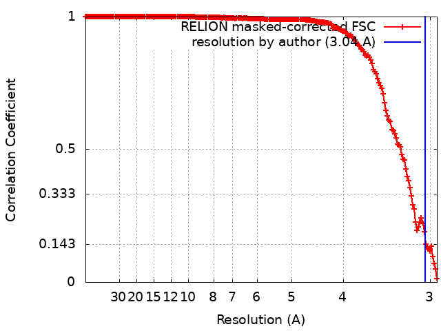 |